Cardiac Evaluations in the Pre-Purchase Exam: How the Echocardiogram Helps Assess Prognosis
F. ter Woort
Learning Objectives
Provide valuable information to your client based on accurate cardiac auscultation and understand the common and uncommon findings on the echocardiogram and what they mean for the horse’s athletic prognosis.
Cardiac murmurs are common in equine athletes, and in a large proportion of cases, these murmurs are compatible with performance at the highest level. The catch, however, is that the intensity of the murmur doesn’t correlate well with the severity of the disease. For example, mitral regurgitation due to mitral valve prolapse, a benign condition with a very good athletic prognosis, can have the same intensity murmur as mitral regurgitation due to degenerative valve disease with a dilated left atrium and a more reserved long-term athletic prognosis. Although over 90% of horses presented for cardiac evaluation as part of their pre-purchase evaluation (PPE) have mild heart disease unlikely to affect their athletic career, a small proportion of horses have severe performance-limiting and occasionally life-threatening cardiac conditions found on their PPE.
As such, a complete cardiac evaluation in any horse with an abnormality on auscultation provides a unique opportunity to remove at-risk horses from the sport. Thankfully, the differential diagnosis for cardiac diseases in horses is relatively straightforward. A thorough cardiac auscultation in a quiet setting provides the diagnosis in the majority of cases.
fig
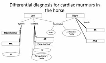
Abbreviations
AR – aortic regurgitation
MR – mitral regurgitation
PDA – persistent ductus arteriosus
PR – pulmonic regurgitation
PS – pulmonic stenosis
TR – tricuspid regurgitation
VSD – ventricular septal defect
In bold square shapes are the common abnormalities, in smaller circular shapes are the much less common abnormalities.
An ACVIM/ECEIM consensus statement was recently published concerning recommendations for equine athletes with cardiovascular abnormalities;1 this is an extremely useful guide when assessing athletic prognosis based on the echocardiography findings.
Mitral Regurgitation
Left-sided systolic murmur over the aortic to mitral valve area: mitral regurgitation.
Why Recommend the Echocardiogram?
Determine the cause if possible because this affects prognosis. Although in many cases there are no specific abnormalities of the mitral valve, in certain cases you can find mitral valve prolapse, which has a good prognosis; a ruptured cordae tendineae or endocarditis, which have a guarded to poor prognosis; or degenerative valve disease, for which the prognosis depends on the extent of the valve thickening and the age of the horse.
Determine the cardiac dimensions: left atrial enlargement will be the first change noted with advancing mitral regurgitation. In severe cases, left ventricular volume overload and pulmonary hypertension can also occur. Horses with normal cardiac dimensions, even with a medium-sized jet of mitral regurgitation are unlikely to be affected by this cardiac pathology.
Determine the size and number of jets: this assessment can be grossly over- or under-estimated based on the Doppler capacities of your portable machine and the Doppler scale settings, and prudence should be exerted when interpreting this parameter. However, finding multiple larger jets in a young horse, even with normal cardiac dimensions, raises some concerns about the possible progression of this condition over time.
What Is the Risk with MR?
Enlarged left atrium is a risk factor for atrial fibrillation, which can affect performance at the highest level. Although many horses in atrial fibrillation are able to perform at lower and medium levels (and some even high levels of dressage and show jumping), they have been shown to exhibit ventricular arrhythmias during exercise,2 raising concern for their safety and that of their rider.
Severe mitral regurgitation can be a primary cause of poor performance.
An exercising electrocardiogram (ECG) is recommended in horses presented for PPE with moderate or severe MR.
Aortic Regurgitation
Left-sided diastolic murmur over the aortic valve: aortic regurgitation.
Why Recommend the Echocardiogram?
Determine the cause because this affects prognosis. Degenerative valve disease of the aortic valve is the most common lesion found in horses with aortic regurgitation over 15 years of age. In these cases, the aortic regurgitation typically progresses slowly. Finding a torn leaflet, endocarditis or valve scarring is associated with a more rapid progression and thus a more guarded prognosis.
Determine the cardiac dimensions: left ventricular enlargement is the first change noted with advancing aortic regurgitation. In severe cases, left atrial enlargement and concurrent MR can also occur. The cardiac dimensions are a major factor in assessing the severity of the AR (much more than the size of the jet).
What Is the Risk with AR?
Horses with AR are at an increased risk for ventricular arrhythmias,3 and sudden cardiac death due to fatal ventricular arrhythmias has been observed in horses with moderate to severe AR1. Severe aortic regurgitation can be a primary cause of poor performance.
An exercising ECG is recommended in horses presented for PPE with moderate or severe AR.
Tricuspid Regurgitation
Right-sided systolic murmur over the tricuspid valve: without concurrent murmurs, this is tricuspid regurgitation. With a concurrent slightly less loud systolic murmur over the pulmonic valve area, this is a ventricular septal defect.
Tricuspid regurgitation is common in equine athletes and typically very well tolerated.
Why Recommend the Echocardiogram?
Rule out endocarditis, especially in a horse with a history of a jugular vein catheter, which has a more guarded prognosis than TR with a normal valve.
Determine cardiac dimensions: use right-sided enlargement to determine severity.
What Is the Risk with TR?
Unless the TR is caused by endocarditis or the right side is significantly enlarged, TR is well tolerated. Significant right-sided atrial enlargement also raises a concern for the development of atrial fibrillation.
Ventricular Septal Defect
Why Recommend the Echocardiogram?
Determine the size of the VSD and the shunt velocity: a defect of 2.5 cm or smaller and a shunt velocity above 4 m/s have a good prognosis, whereas a defect larger than 2.5 cm and shunt velocities under 4 m/s are more likely to have hemodynamic consequences.1
Determine cardiac dimensions: Left-sided enlargement is the first sign of the effect of over-circulation of the pulmonary vasculature induced by the VSD. Advanced cases can show pulmonary hypertension and congestive heart failure.
Determine the presence of concurrent abnormalities: due to its most common location in the septum just below the aorta, the aortic valve can prolapse into the defect. This initially renders a functionally smaller defect, but destabilizes the aortic valve leading to aortic regurgitation, which worsens the left ventricular volume overload. Left-sided enlargement can lead to MR, further worsening the left atrial volume overload.
The VSD can be a component of other more complex congenital abnormalities.
What Is the Risk with a VSD?
Large VSDs can lead to poor performance. Ventricular arrhythmias and atrial fibrillation have appeared in horses with VSDs for which the risks were detailed above. An exercising ECG is recommended in horses presented for PPE with a VSD.
Other Abnormalities Less Commonly Encountered
A continuous machinery murmur on the right side of the chest: aortocardiac fistula—these horses are unsafe to ride and this condition is life threatening. The reason to recommend the echocardiogram is to confirm the diagnosis before condemning the horse (for example concurrent tricuspid regurgitation and very loud aortic regurgitation could produce a somewhat similar sound on the right).
Any diastolic murmur in a Friesian should prompt an echocardiogram: these horses are prone to aortopulmonary fistulas which are incompatible with athletic use and life threatening.
Other abnormalities which can significantly affect performance, encountered on the echocardiogram without an associated murmur are myocarditis and cardiomyopathy. Although these are typically thought to induce clinical signs that would preclude these horses from being presented to a PPE, a few of these cases have been detected on PPE, again underscoring the important role of the echocardiogram in removing at-risk horses from the sport.
References
1. Reef VB, Bonagura J, Buhl R, McGurrin MKJ, Schwarzwald CC, van Loon G, et al. Recommendations for management of equine athletes with cardiovascular abnormalities. J Vet Intern Med. 2014;28:749–61.
2. Verheyen T, Decloedt A, van der Vekens N, Sys S, De Clercq D, van Loon G. Ventricular response during lungeing exercise in horses with lone atrial fibrillation. Equine Vet J. 2013;45(3):309–14.
3. Marr C. Cardiac murmurs: acquired valvular disease. In: Marr C, Bowen M, editors. Cardiology of the Horse. Saunders; 2010:207–16.