Introduction
Primary CNS lymphoma was extremely rare and that accounted for only 4% of all primary intracranial tumor. In canine, differentiation of lymphoma subtypes can be performed by histopathological evaluation and immunophenotyping. Subtyping is crucial for prognosis and therapeutic plan.
Objectives
We describe clinical findings, management, and pathological diagnosis of a dog with CNS lymphoma. Subtyping of the tumor was done by immunohistochemistry for CD3 and CD79a.
Methods
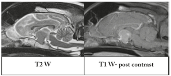
| Figure 1 | 
Sagittal MR imaging mass effect at extraparenchymal structures on sellar region which were hyperintense on T2-weighted images and contrast enhancement on T1-weighted images. |
|
| |
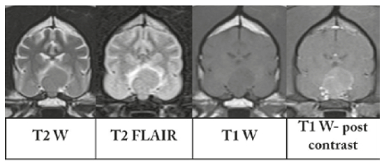
| Figure 2 | 
Transverse MR imaging of brain showed hyperintense on T2-weighted images, perilesional hyperintensity on FLAIR images, hypointense on T1-weighted, and contrast enhancement on T1-weighted images. |
|
| |
An eight-year-old, intact male crossbred dog was presented neurological deficits with disoriented, left circling, nystagmus, and left cranial nerve deficits. Blood profile was within normal limits. Superficial lymph nodes appeared normal at palpation. Magnetic resonance imaging identified mass effect at extraparenchymal structures on sellar region nearby the pituitary gland which were hyperintense on T2-weighted images, contrast enhancement on T1-weighted images and had perilesional hyperintensity on FLAIR images. Prednisolone was used as an anti-inflammatory drug and after that clinical sign was improved to conscious and circling was not detected. Thirty days later neurological signs progressively worsened, obtunded, inappetence, respiratory distress, and the dog was died from respiratory arrest.
Results
Gross examination revealed that the mass was located at sellar region of the brain. Histopathological evaluation revealed aggregated round cells and lymphocytes in this region. The round cells had moderate amounts of cytoplasm, finely stripped nucleus, and some of these cells were bi-nucleated with nuclear molding. Immunohistochemical analysis was performed for CD3 and CD79a, and the tumor was characterized as T-cell-rich B-cell lymphoma.
Conclusions
We reported a rare case of primary CNS lymphoma, subtyping T-cell-rich B-cell lymphoma.