Principles and Clinical Application of Cerclage Wiring
Orthopedic wire is used as a supplementation for fracture fixation for the reduction of fragments and the protection of fissures. Never use wire as a sole means of fracture repair. It is most commonly used as an adjunct to intramedullary (IM) pin fixation. The degree to which this device helps to neutralize the remaining forces acting on the fracture site also is directly dependent upon the fracture configuration; for example, full cerclage wires effectively neutralize rotational, compressive and shear forces in a long oblique fracture - but are ineffective in a short oblique or transverse fracture.
Implants
Orthopedic wire is provided as a malleable form of 316L stainless steel. It comes in a range of diameter, from 0.5 to 1.5 mm (2416 gauge). The two types of orthopedic wire available are gauge wire and loop wire. Gauge wire comes in spools or long strips (coil type). Loop wires have a preformed eye at one end for a single knot and are available in 0.85, 1.0, 1.25, and 1.5 mm diameters and in 150 and 300 mm length.
Tightening Methods
The three most common methods for tightening and securing cerclage are twist, single loop, and double loop knots.
Twist Knot (a)
To form a twist knot, old needle forceps or pliers can be used. Instruments that lock the two strands may help ensure that the twist forms with an even wrap. The first two to three twists should be formed by hand. The loose twist is then grasped with the instrument and further twists formed while pulling very firmly away from the bone. This will ensure that the twist is tightened at its base, and that the wires wrap around each other, not one around the other. Once the wire is tight (meaning that doesn’t longer move if pushed) or the surgeon feels that further twist will cause the wire to break, the twist is cut leaving 3 wraps of wire. It is very important not to wiggle the cerclage wire while cutting it, otherwise there is the risk of undoing the twist and loose the tension. Another important recommendation is to not push the twisted wire down to adhere to the bone surface as this maneuver will generally reduce the initial tension.
Single Loop Knot (b)
Single loop knot is formed using a length of wire with an eye at one end. The free end is passed around the bone and through the eye, and tip of the tightener. It is then passed into the wire tightener rotating crank. The wire is tightened around the crank, and by rotating it the wire is tightened. Once the desired tension is achieved, the wire tightener is bent over to lock the free end within the eye. The wire tightener is loosened and the free end pushed down so that it is bent back completely on itself. The wire is then cut, and flattened further if needed.
Double Loop Knot (c)
Double loop knot is formed using a length of wire that is folded over on itself to present a double strand of wire. The fold is compressed but left open enough to be able to pass two strands of wire through the bent end. The folded end is passed around the bone, and the two free ends passed through the loop. The wire is pulled tight. The free ends then are passed into the wire tightener using 2 cranks. Each wire end is loaded into one of the cranks in the same manner as for single loop cerclage. The two cranks are tightened simultaneously until the tension “feels” appropriate, and the wire tightener is bent over, again while maintaining tension in the cranks. Once the bends are sufficient, the cranks are loosened, and the bending completed so that the wire ends lie flat to the bone. The wires are cut, and the arms flattened further if needed.
Clinical Considerations
Full Cerclage Wiring
Full cerclage wiring is when the wire is placed around the bone. This technique is used for long spiral and oblique fractures and to protect fissures. The bone must be completely reconstructable for this technique to work. General “rules” for full cerclage wire application follow:
- The length of the fracture line obliquity should be at least twice the diameter of the bone.
- All wires should be spaced approximately 1.0 cm apart, and all wires should be placed at least 0.5 cm from the end of a fracture.
- All wires need to lie directly on bone (no interposition of soft tissues).
- All wires must be placed perpendicular to long axis of the bone.
- All wires should be placed and tightened before the primary fixation applied.
Hemi-cerclage Wiring (Inter-fragmentary Wiring)
This is used to counteract forces of rotation and shear in transverse and short oblique fracture. Drill holes in each bone segment and pass the wire through the holes. This technique adds little to the mechanical stability of a fracture. The holes in the bone weaken the fragment ends. Inter-fragmentary wiring is useful for repair of some mandibular.
Principles and Clinical Applications of Tension Band Wiring
Tension band wiring is a means of converting the distractive forces acting at the fracture site into compressive forces between the fragments. Such distractive forces are generally caused by a tendon or ligament being attached to an avulsed bone fragment. Conversion of these forces to create compression at the fracture surfaces will generate stability and lead to bony union.
Technique
Avulsion fragments can be effectively stabilized using the pin and tension band wire technique.
The fragment is initially stabilized using either 1 or 2 K-wires. To protect this implant from being pulled out or bent by the forces exerted by the pull of the attached ligaments or tendons, a cerclage wire is placed to oppose the tensile forces. An additional and very significant benefit of this technique is that the pull in the ligament and the counter-pull in the wire convert these tensile forces to compressive forces across the fracture.
In principle, the use of two small parallel pins is better than a single bigger pin; however, the number and size of pins will be defined by the size of the fragment.
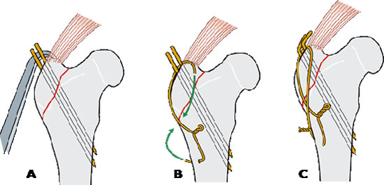
Once the fragment is reduced, the pin(s) are driven perpendicular to the fracture plane and parallel to each other. If possible, they should penetrate the trans-cortex (Figure 1A). To anchor the tension band wire, a hole is drilled transversely across the main fragment on the surface away from the ligament or tendon. This hole is usually located approximately the same distance below the fracture line as the pins are above the fracture line. It is important to ensure when drilling the hole that sufficient bone is purchased to prevent the wire from cutting through it and causing a ‘stress riser’ point that could potentially result in a pathological fracture.
The wire is passed through the hole in the bone, brought across to the original side of the bone, around the ends of the pins and back to the other end of the wire on the starting side of the bone. This creates a figure-of-eight pattern (Figure 1B).
Twist knots are commonly used for tension band wires. To effectively tighten a tension band wire with one knot, the slack in the arm opposite the one with the knot must also draw in by the tightening process. This is very difficult in bigger wire sizes. To avoid this, a twist knot can be tied in both arms of the figure of eight.
Once the wire is tightened, the ends of the K-wire are bent over so that they lie flat against the bone (Figure 1C).
| Figures 1A,B,C | 
|
|
| |
Clinical Applications
The main indications for tension band wiring is the treatment of avulsion fractures such as those involving the olecranon, greater trochanter, patella, tibial tuberosity malleoli, calcaneus.
In all these avulsion fractures, the fragment is distracted by the muscle, tendon or ligament which originates/inserts on it.
The tension band is placed so that counteracts the tensile forces acting on the fragments and redirects it to compress the fragment against the adjacent bone. Tension band wiring is also used to repair osteotomies, for example trans-olecranon osteotomy in the caudal approach to the elbow joint or osteotomy of the greater trochanter in the dorsal approach to the hip joint.
Case Example
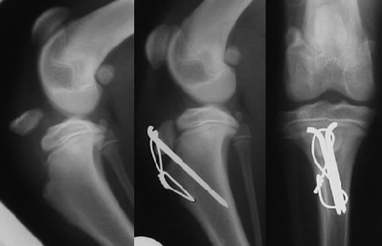
The treatment of an avulsion fracture of the tibial tuberosity is used here as an example of tension band wiring (Figure 2). This injury is quite common in greyhounds and terrier breeds and the fracture occurs through the growth plate. The tibial tuberosity is distracted by the tensile forces exerted by the quadriceps muscle group through the straight patellar tendon.
The fracture is initially reduced and fixation is achieved with two K wires driven through the tuberosity into the metaphysis. The idea behind placing two K wires is that they supposedly prevent rotation of the fragment. In reality, a well-reduced avulsed fragment attached to the tendon has very little tendency to rotate, particularly once it is under compression, therefore a single pin is in most of cases more than adequate for the purpose. If the choice lies between one large and two smaller pins, the latter is the best choice. A hoe is drilled transversely through the tibia distal to the fracture site.
| Figure 2 | 
|
|
| |
A length of orthopedic wire is passed through the hole; it is very important to pay attention to use the correct diameter of the wire. As a general indication:
- For patients over 20 kg of bodyweight use 18 gauge (1.2 mm diameter)
- For patients below 20 kg of bodyweight use 20 gauge (1.0 mm diameter)
The ends of the wire are brought across the cranial aspect of the tibia in a “figure of 8” pattern and then passed under the straight patellar tendon before being twisted tight. As for cerclage wire twisting technique, it is important to ensure that the two wire ends are correctly twisted around each other and not one around the other to form a “skip knot.”
As the wire is tightened, its proximal loop engages on the protruding ends of the Kirschner wires. Each K wire is then bent and cut leaving a hook of about 5 mm long that is rotated to fit snugly against the tibial tuberosity and insertion of the straight patellar tendon.
Although theoretically a twist should be placed in each side of the “figure of eight” to achieve uniform tension in the wire, adequate tightening is achieved with a single twist on just one side, particularly in short tension band constructs.
References
1. Roe SC. Mechanical comparison of cerclage wires: introduction of the double-wrap/loop and loop/twist types. Vet Surg. 1997;26:310–316.
2. MacLaughlin RM. Internal fixation: intramedullary pins, cerclage wires and interlocking nails. Vet Clin North Am Small Anim Pract. 1999;29:1097–1116.
3. Johnson AL, Houlton JEF, Vannini R. AO Principles of Fracture Management in the Dog and Cat. Thieme - 2005 by AO Publishing, Switzerland, Clavadelerstrasse, CH-7270 Davos Platz. Distribution by Georg Thieme Verlag, R.