Prof. Dušan Palić, DVM, MVSc, PhD, CertAqV, DECAAH
Faculty of Veterinary Medicine, Ludwig-Maximilians University Munich, Munich, Germany
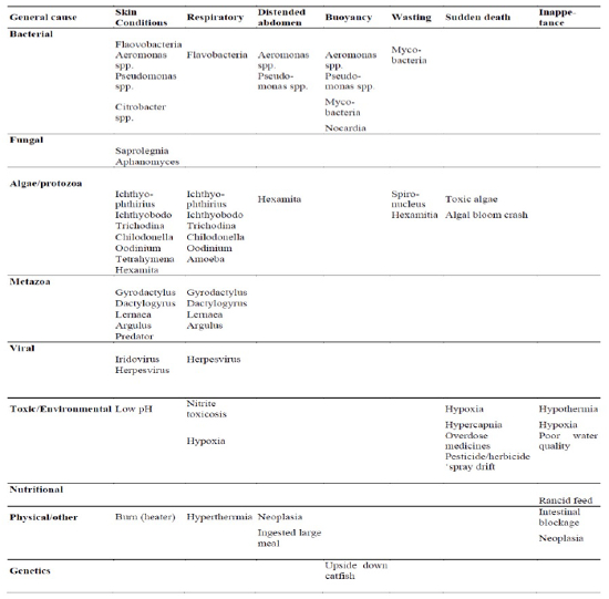
Fishes show a range of clinical signs when diseased. What are these, and what do they mean? There are clinical signs that are pathognomonic for certain diseases, however, many are non-specific and a step-by-step approach to working up a case is necessary. First, it is very important to familiarize yourself with what is normal in terms of appearance and behavior for the species you are dealing with. Typically, healthy fish should have a good appetite; have clean, clear, vibrant body coloration; hold their fins erect; and have bright red gills. They should be active and not display abnormal behavior, swimming patterns, or have loss of buoyancy control. However, there are always exceptions to these rules as the types of different fish species number in the thousands. There are some fishes such as the wrasses that lie on the bottom or on their side, there are upside down catfish that swim upside down and there are fish (goldfish) with deformities which are all ‘normal’. In the following paragraphs, I will describe pathological presentations and how they may be interpreted in terms of pathobiology and etiology. Further down, there is a table of common etiologies as they relate to the general syndromes that are discussed (Table 1).
Table 1. Common clinical signs in fish - a review (adapted from Loh and Landes)
Skin conditions can vary from erosions on the mouth or fins (usually due to Flavobacterium infection) or along their lateral line system (e.g., hole-in-the-head disease). Ulcers are common and they can originate from the outside (e.g., Aeromonas spp.), or be ulcers that originate from the inside (mycobacterial). Ulcers may present as discrete lesions anywhere on the body including the fins, flank, and operculum. They can be circumscribed and show advancing border. They may be pale to red, depending on the depth of the ulcer. The pale ulcers are more superficial and indicate edema and the red may be due to hyperemia. A dull red appearance is evidence of deeper ulceration with exposed muscle. Such deep ulcers may be due to fungi (e.g., Aphanomyces invadans), protozoa (e.g., Tetrahymena), or simply from a predator attack. It is more common that the skin ulcers are due to secondary bacterial infection, with the primary pathogens being either skin flukes or lice. Thus, it is very important to investigate for the primary cause. Sometimes, melanization occurs in response to injury which is commonly seen in goldfish.
Fish may have proliferative skin lesions that may be raised and smooth (e.g., carp pox and neoplastic conditions) or be fine and granular (e.g., lymphocystis). They may present with fine white spots (e.g., white spot disease) or appear as a haze (e.g., velvet disease) or larger spots (e.g., digenetic trematodes). Excessive slime production may be an indication of ectoparasitism or poor water quality issues (e.g., low pH). A change in body color patterns, whether it be pale or dark is nonspecific. Fish may have tuft-like white growths which can be due to fungi (Saprolegnia) or bacteria (Flavobacteria). Hyperemia of fins or body is a common sign of stress and/or bacterial infection. Often, fish with skin lesions may present with flashing (scraping against substrate or tank walls), have clamped fins and separate from the group. If they are infested with particularly irritant parasites (e.g., Argulus), the fish may jump in an attempt to dislodge the parasites. Generally, those with severe disease may become lethargic and very often display respiratory signs of disease too. The reason for this is because the gills are also in intimate contact with the water and the external environment. Thus, many pathogenic organisms that colonize the skin will also affect the gills.
Respiration: It is always a good idea to check that the gill color is a healthy bright red. Pale gills indicate anemia, whereas dark gills indicate methemoglobin formation. Gills with excessive mucus indicate ectoparasitism or dissolved toxin. Whenever gills are damaged, they have a limited range of response and they include formation of synechiae (secondary lamellae that ‘stick’ to each other), epithelial hyperplasia, secondary lamellar fusion and if given sufficient time, mucus cell hyperplasia. All these will decrease the efficiency of gill function and fish will display respiratory signs of disease. Fish may congregate at water inlets and ‘pipe’ or ‘gasp’ at the water surface. The opercular beat rate may initially be increased as the fish try to respire through inefficient gills, but as fish become moribund, the opercular rate will decrease.
Under the heading of ‘distended abdomen’ we have proliferative conditions (ovarian neoplasia is common in koi) or cystic conditions (polycystic kidney disease common in goldfish), ‘bloat’ and ‘dropsy’. Bloat is typical for certain cichlid fish known as ‘Malawi bloat’ which is caused by Hexamita (an intestinal flagellated protozoa). But enteric infections with bacteria such as Pseudomonas can also present in a similar fashion. The common term of ‘dropsy’ is used when there is also protrusion of the scales to create a ‘pine cone’ appearance due to subcutaneous edema. This is obvious in fish with larger scales, but is difficult to appreciate in species such as the angelfish with fine scales. Dropsy is often accompanied by ‘pop-eye’ (exophthalmia). These are commonly a result of primary or secondary bacterial infections causing inflammation and vascular damage, especially to the rich vascular beds in the kidney and choroid rete behind the eye. The insult to the kidney interferes with fluid balance, causing the ‘dropsy’ appearance and inflammation behind the eye causes the ‘pop-eye’ appearance. There is one breed of goldfish where exophthalmia is selected and these are known as telescope moors.
Fish in advanced disease can present with buoyancy disorders. They may either become negatively buoyant and sink to the tank floor, or become positively buoyant, floating to the surface. This is a very common condition in goldfish breeds and in my experience, it is more common in rotund breeds such as the Ryukin, Pearlscale, and Orandas. These fish tend to also have twin tails. There is suggestion that fish should not be overfed and that they should be given adequate fiber in their diet. Systemic bacterial infections are also common causes of buoyancy disorders. Less common causes include coccidial and fungal infections of the swim bladder.
Fish that are wasting present with concave abdomen. The differential diagnoses for poor body condition in fishes include, but are not confined to the following: chronic malnutrition, or infections by bacteria (e.g., mycobacteriosis), protozoal organisms (e.g., Hexamita, Spironucleus, Cryptobia, Sporozoa, lchthyobodo) and metazoa (e.g., Gyrodactylus, Dactylogyrus, and a number of cestode species). Fish with enteropathy will have long fecal strings, that may contain bubbles and float, or they may be empty fecal strings (in most freshwater fishes, normal fecal casts should resemble black string). If they are overfed, their fecal strings take on the same color of the fish food fed to them. Those with enteritis will display congested vents. This can be overlooked when assessing health of pond fish. Thus, it is very important to capture some fish to examine their vents.
Causes for sudden death are difficult to diagnose because fish tissues autolyse rapidly. Based on epidemiological principles, sudden death is likely due to environmental causes. Collating a good history and water quality analyses are important.
Inappetence is a non-specific sign of illness. It can also occur when water temperature deviates from their tolerance range. The most common is during winter when owners are unaware that they need heating for their Siamese fighting fish, or their heater has stopped working. You will see remains of uneaten food in the tank or filter.
So, we have discussed the clinical signs displayed by fish during ill-health. But some other things we should also consider include their environment. Fish that are sick tend to produce more mucus and you may notice a fishy smell coming from the tank and perhaps there may be excess stable foam accumulating at the water surface.
References
1. Loh R, Landos M. Fish Vetting Essentials. Perth: Richmond Loh Publishing; 2011.
2. Roberts H, ed. Fundamentals of Ornamental Fish Health. Ames, IA: Wiley-Blackwell; 2010.
3. Noga, EJ. Fish Disease: Diagnosis and Treatment, 2nd ed. St. Louis, MO: Mosby; 2010.