Bilateral Ovarian Neoplasia Mimicking Dysplastic Kidneys in Ultrasound Imaging
A. Lobaczewski1; 0. Szalus-Jordanow2; M. Czopowicz3; A. Kosiec-Tworus4; A. Olkowski1; T. Frymus2
Introduction
An intact, female Yorkshire Terrier, aged 14 years, was referred to the clinic due to lack of appetite and dyspnea. On admission, the body temperature was 38,9°C, and besides dyspnea abdominal distension was seen.
Objectives
To determine the cause of the poor clinical state of the animal.
Methods
Ultrasound and X-ray examination. Surgical treatment was applied and histopathological examination of the ovaries was performed as well as microbiological examination of the abdominal fluid.
Results
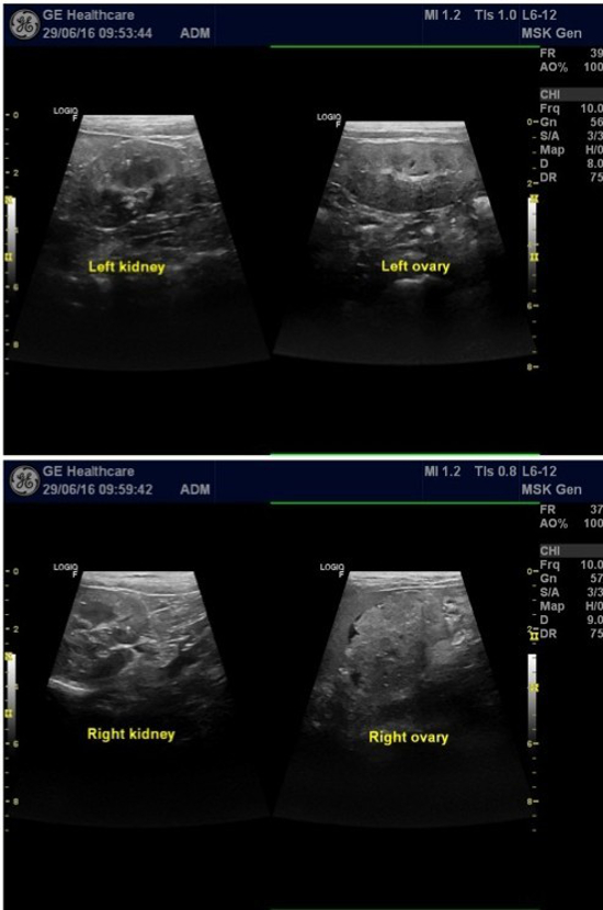
In the ultrasound examination on both sides caudally to the kidneys (in a typical location for ovaries) solid, oval masses, sized 2,5 cm x 4,4 cm, with mixed echostructure and visible capsules were seen (Figure 1). Peritoneal fluid with significant cell content was also confirmed. An exploratory laparotomy revealed that both ovaries were enlarged. Free peritoneal fluid was removed and then the ovaries and uterus. Histopathological examination of the ovaries revealed an adenoma or low grade carcinoma . For several weeks post-surgery the clinical state of the patient was good, and ultrasound as well as X-ray examination did not show metastases. However, 4,5 months from the surgery the dog died showing symptoms of severe dyspnea and circulatory collapse. A necropsy was not permitted.
| Figure 1 | 
|
|
| |
Conclusions
That is a case of a very rare ultrasound image in that tumors of the ovaries looked like dysplastic kidneys. Our 2 ultrasonography specialists (more than 10 years of experience each) have seen similar images only three times.