Concentric Left Ventricular Hypertrophy in a Dog Secondary to a Pheochromocytoma and Systemic Hypertension - Case Report
O. Szalus-Jordanow1; A. Lobaczewski2; R. Sapierzynski3, M. Czopowicz4; T. Frymus1
Introduction
A 12-year-old female Scottish terrier was referred to the clinic because of lethargy, lack of appetite and occasional vomiting. The animal poorly responded to previous supportive treatment. On admission, the dog was unconscious and dehydrated.
Objectives
To determine the cause of the poor clinical state of the animal.
Methods
Ultrasound and echocardiography examination, blood pressure measurement and necropsy.
Results
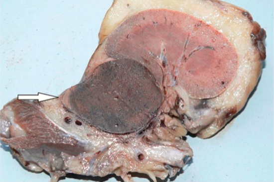
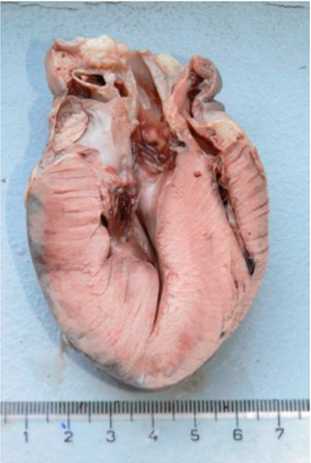
Ultrasound examination revealed a 31x45 mm tumor of the left adrenal gland and an oblong 37x1.1 mm mass in the lumen of the caudal vena cava. Echocardiography showed massive concentric left ventricular (LV) hypertrophy (thickness of the intraventricular septum and of the left free wall was 17 mm and 13 mm, respectively). Systolic arterial blood pressure measured by oscillometry was 189 mm Hg, diastolic - 132 mm Hg, and mean - 151 mm Hg. The dog died soon after admission. Autopsy confirmed an adrenal tumor (Figure 1), a caudal vena cava thrombus and LV hypertrophy (Figure 2).
| Figure 1. Pheochromocytoma | 
|
|
| |
| Figure 2. Left ventricular hypertrophy | 
|
|
| |
Microscopically the tumor was a pheochromocytoma, as confirmed by immunohistochemistry with antibodies against synaptophysin. Microscopic examination of the heart revealed multifocal area of mild fibrosis accompanied by inflammatory infiltration with predominating small lymphocytes. Multifocal hypertrophy of cardiomyocytes characterized by the presence of enlarged vesicular nuclei with visible nucleoli and abundant sarcoplasm was also observed. In some of these cells brown granules of lipofuscin were also present.
Conclusions
This is a rare case of LV hypertrophy in dogs caused by a pheochromocytoma and systemic hypertension.