Introduction, Clinical Investigation and Force Plate of Patients with Elbow Dysplasia (ED)
Department of Clinical Sciences of Companion Animals, Utrecht University, Utrecht, Netherlands
Introduction
Originally the term "elbow dysplasia" or "dysplasia articulationis cubiti" covered generalized osteoarthrosis of the elbow joint with an ununited anconeal process (UAP) (Corley et al. 1968). These days UAP is just one of the different entities which are covered by the term "elbow dysplasia" which is here defined as the group of elbow dysplasias including: 1) ununited anconeal process (UAP), 2) fragmented medial coronoid process (FCP), 3) osteochondrosis (OC) or osteochondritis dissecans (OCD), and 4) incongruity (INC) of the elbow joint. These four entities have in common that they all occur in the elbow joint (although OCD occurs also in other joints), that they are all seen in young growing dogs of 4–6 months of age (although they can be overlooked and reveal the first signs at mature age) of medium and large size, that they can cause lameness (but not in all cases these entities go together with lameness, and if so not always for the first time at young age), and that they will cause osteoarthritis (OA) (but that can vary per individual dog and perhaps even per breed). Since developmental skeletal diseases, either due to genetic cause or due to nutritional influences or trauma, are frequently seen in this category of companion animals all three etiologies can be held responsible for the occurrence of ED. Here, first definitions and a short indication of its etiology will be given of each of the primary entities of ED.
Primary Lesions in the Screening Programme According to the International Elbow Working Group (IEWG)
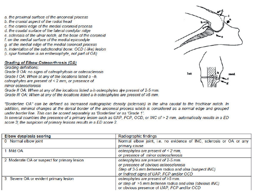
These lesions are graded as absent (ED grade 0), suspected-present (ED grade 2) or obvious present (ED grade 3) (Figure 1). We will distinguish the following primary lesions:
1) Ununited Anconeal Process (UAP)
Separation in the cartilaginous bridge between the secondary ossification centre of the anconeal process and the olecranon, which can cause a partially or completely detached anconeal process, is referred to as ununited anconeal process.
Etiology: When the humeral condyle increases in proportion (i.e., in diameter), the semilunar notch should widen equally by shifting the anconeal process in a proximal direction. However, when widening stays behind or at the same time the radius pushes the humerus in a proximal direction (in case of delayed ulnar growth in length) this can lead to a shift of the anconeal process off its origin when (i) the anconeal process is a secondary ossification centre (as it is in some but not all breeds) and (ii) it is not bony fused yet with the ulna (i.e., at the age of < 5.5 months).
2) Fragmented Medial Coronoid Process, Medial Coronoid Process Disease (MCPD), and Medial Coronoid Disease or Medial Compartment Disease (MCD)
Fissuring of the medial coronoid process of the ulna with partial to complete separation (fragmentation) of the medial coronoid process from the ulna.
Etiology: Primary osteochondrosis of the subchondral bone with a fissure line between osteochondral cartilage and subchondral bone, possibly with secondary a fissure line in overlying articular cartilage as described by Guthrie (1992) and recently by Lau (2014, and Lau: this proceedings), although also chondromalacia at the medial coronoid process is considered part of this entity.
3) Osteochondrosis or Osteochondritis Dissecans
Local thickening of growing epiphyseal cartilage of the distal humerus due to delayed endochondral ossification (i.e., osteochondrosis = OC), which may develop into a single or fragmented detached cartilage flap (i.e., osteochondritis dissecans = OCD).
"Kissing lesion": An abrasion of the articular cartilage of the humerus, sometimes extending into the subchondral bone (and radiological often slightly more lateral than the OC-lesion), is caused by a fragmented coronoid process at the opposite side as suggested by Morgan in 2000. This finding is graded as a "OCD-like lesion" since it is not always possible to distinguish it from OC/OCD (both in Fig. 1 at location "h").4
4) Elbow Incongruity (EI, INC)
The subchondral bone of the trochlear notch of the ulna and of the radial head are not parallel to the opposing humeral subchondral bone.
There are different forms of EI:
i. The radius is longer than the ulna with a narrowing of the joint space between the tip of the anconeal process and the humeral condyle, a distally gradual widening of the joint space between the ulnar semilunar notch and the humeral condyle and the radial head proximal of the coronoid process of the ulna (this form of EI is typically seen in Bernese Mountain dogs, often together with a fragmented apex of the coronoid process (FCP).
ii. The longer ulna with a wider joint space between the proximal radius and the humeral condyle and the step between the more proximally located distal edge of the ulnar trochlear notch (i.e., the lateral coronoid process) and the radial head (and displacement of the distal humerus cranially). This can also be considered as an underdeveloped or too small trochlear notch (or part of traumatic short radius syndrome).
iii. The alignment between the subchondral bone of the trochlear notch and the radial head is more elliptical than the circular contour of the humeral condyles described by Wind, especially in German Shepherd Dogs, in 1986.
iv. An incongruity of the radial-ulnar joint (not to be detected on plain radiographs) warrants computed tomography, but will only visualize the subchondral bone; this incongruity may play a role in avulsing the loosening of the medial coronoid process when weakened. Developmental elbow luxation with lateral displacement of the (often hypoplastic) radial head with a comparative overgrowth of the radius (as seen in chondro-dysplasia in non-chondrodystrophic breeds) can be described as elbow incongruity although it is generally considered beyond the scope of the screening for ED.5
Osteoarthrosis (OA) is radiologically characterized by new bone formation at the edges of the joint cartilage. In addition, enthesophytes (i.e., new bone formation at the sites of attachments of tendons, ligaments, and joint capsule, resulting from abnormal tension placed on the soft tissue attachments near, but outside the joint) can be formed; quite typical is the enthesophyte at the caudal margin of the humeral condyle (Fig. 1, indicated by "i ") in case of more severe ED. Regardless of the primary cause, the pattern of OA is similar. The different locations where osteophytes and enthesophytes are visible in case of OA are given in Fig. 1 (a, b, d, f, g, and i). Both secondary signs (OA) and primary signs (UAP, FCP, OCD, INC) play a role in the final scoring for ED (Fig. 1). An irregularity at the dorsal margin of the anconeal process (in Fig. 1 indicated with "a"), can be physiological in certain breeds (Lappalainen 2014 in: http://www.vet-iewg.org/ under proceedings 20146).
Sclerosis, an alteration in normal bone architecture (i.e., a decrease in normal bone porosity) is depicted on a ML view of the elbow joint as an increase on bony opacity with loss of trabecular markings (a white area), within the trochlear notch just caudal to the lateral coronoid process (Fig.1 at location "e"). Osteosclerosis is considered as one of the first signs of ED in young dogs, especially when the primary cause cannot be identified as in some cases of FCP. Since FCP often occurs bilaterally, the use of the opposite elbow joint for comparison will not always be of help to detect sclerosis. In a survey with 17 Labrador Retrievers (6–16 months of age) with FCP and 17 without FCP as diagnosed by arthroscopy, radiographic density was objectively diagnosed and expressed as pixels: an extremely significant correlation between pixel intensity of the projection of the lateral coronoid process revealed in dogs with FCP (Burton et al. 2007). Microscopically, this area is characterised by reduced intertrabecular spaces (Wolschijn et al. 2004) in the medullary cavity of the ulna, either due to mechanical overloading or influence of e.g., MMPs, enzymes which play a role in OA (Fig. 1).
| Figure 1. Locations for grading of elbow OA (http://www.vet-iewg.org/) | 
|
|
| |
Clinical Investigation
It is of paramount importance to see the dog walking and trotting on a loose leash in a quiet surrounding before deciding at which leg the dog is (the most) lame. Then the dog is put on the table and investigated (looking and superficial palpation) from proximal to distal comparing contours of muscles, bones and joints. On lateral recumbence the leg is meticulously investigated from distal to proximal (from nails to cartilage scapulae) with special attention for all joints (range of motion, crepitation, pain reaction). The elbow joint is flexed and extended several times with the thumb placed at the anconeal muscle for crepitation and a close look at the reactions of the dog for pain sensations. In addition, pronation and supination is performed in different flexed positions and supination with the elbow extended to check for pain reaction. In addition the shoulder joint is investigated (extension and flexion) and flexion of the shoulder plus extension of the elbow joint (the latter to check the biceps tendon) is performed. Finally deep palpation of the long bones is performed for bone pain (like in panosteitis).
Typical signs of ED are: joint effusion in case of OA (especially UAP), new bone formation at the caudolateral margin of the humeral condyle (in case of OA), pain upon extension (in case of UAP or incomplete humeral fractures) or flexion, supination and pronation at different angles (FCP, OCD), pain upon extension plus supination, decreased range of motion especially in flexion (in case of OA). Based on the clinical findings imaging techniques are to be considered (see presentations of Dr. Boroffka, Dr. Lau, Dr. Heng and Dr. Ondreka, this congress).
Force Plate Technique
Different techniques are in use to investigate in an objective way the locomotion of dogs affected with ED. One of these is the measurement of ground reaction forces. Rather than only evaluating the vertical force (Fz), the advantage of measuring also breaking and propelling ground reaction forces of the front legs give extra insight in the use of each (front) leg. The weight bearing of the dog occurs for 60% by the front legs and the function of the front legs is mainly breaking (Fy-) (rather than propelling = Fy+); consequently, the breaking force and the vertical force are interesting to consider in dogs with front leg lameness. Breaking and propelling forces are unnatural, and thus less informative, when measured at a treadmill (Fanchon & Grandjean 2007) with only a fair to moderate correlation of the data between force plate analysis (FPA) with or without treadmill could be shown in dogs with hind leg lameness (Böddeker et al. 2010). Breaking and propelling forces are totally missed when a force mat is used. The advantage of FPA is that there is only a small variation in repeated measurements allowed to evaluate treatment modalities in case of lameness (e.g., medication, surgery) and that a symmetry index (SI = ratio affected side:contralateral side) is relevant for strictly unilateral pathology (Theyse et al. 2000); the disadvantage is that interpretation can be influenced when bilateral abnormalities are present (as in almost 70% of cases of ED) or when evaluating OA. Therefore, real standardized conditions (body weight, pre-screening exercise, constant walking velocity, one handler) for FPA are of utmost importance. FPA of seven dogs with unilateral FCP was performed before, and 6 weeks and 6 months after, surgical FCP-removal and Fz, Fymax and Fymin were determined for each dog and the SI was calculated (Theyse et al. 2000). The combination of the SIs of Fy-, Fy+ and the impulse of the vertical force (Iz) proved to be more sensitive in determining front leg lameness due to unilateral FCP, than the SI of the maximal vertical force (Fzmax). Fzmax returned to normal values 6 months after surgery for all seven dogs, whereas the other parameters showed persisting abnormalities in two of the seven dogs and the remaining 5/7 dogs had complete normalized SIs, this despite radiological progression of OA in 4/7 dogs at 6 months after surgery (Theyse et al. 2000). This demonstrates that in this study there is no correlation between lameness and the radiological OA-grade, but there is correlation between the lameness and the presence or absence of the fragmented coronoid process.
References
1. Böddeker J, Drüen S, Nolte I, Wefstaedt P. Comparative motion analysis of the canine hind limb during gait on force plate and treadmill. Berl Munch Tierarztl Wochenschr. 2010;123:431–439.
2. Burton NJ, Comerford EJ, Bailey M, Pead MJ, Owen MR. Digital analysis of ulnar trochlear notch sclerosis in Labrador retrievers. J Small Anim Pract. 2007;48:220–224.
3. Fanchon L, Grandjean D. Accuracy of asymmetry indices of ground reaction forces for diagnosis of hind limb lameness in dogs. Am J Vet Res. 2007;68:1089–1094.
4. Hazewinkel HAW, Mei BP, Theyse LFH, Van Rijssen B. Locomotor system. Ch. 17. In: Rijkberk A, van Sluijs FJ, eds. Medical History and Physical Examination in Companion Animals. Edinburgh, UK: Saunders; 2009:135–159.
5. Lau SF, Hazewinkel HA, Grinwis GC, Wolschrijn CF, Siebelt M, Vernooij JC, Voorhout G, Tryfonidou MA. Delayed endochondral ossification in early medial coronoid disease (MCD): a morphological and immunohistochemical evaluation in growing Labrador retrievers. Vet J. 2013;197:731–738.
6. Lau SF, Wolschrijn CF, Hazewinkel HA, Siebelt M, Voorhout G. The early development of medial coronoid disease in growing Labrador retrievers: radiographic, computed tomographic, necropsy and micro-computed tomographic findings. Vet J. 2013;197:724–730.
7. Theyse LFH, Hazewinkel HAW, van den Brom WE. Force plate analysis before and after surgical treatment of unilateral fragmented coronoid process. Vet Comp Orthop Traumatol. 2000;13:135–140.
8. Wolschrijn CF, Weijs WA. Development of the trabecular structure within the ulnar medial coronoid process of young dogs. Anat Rec A Discov Mol Cell Evol Biol. 2004;278:514–519.