Introduction
Porencephaly is an uncommon cerebral lesion secondary to focal neuronal migration disorder or tissue breakdown by several etiologies such as infection, trauma and ischemia.
Objectives
This study aimed to describe the MRI features of an incidental finding compatible with bilateral porencephaly in an asymptomatic mature cat.
Methods
An 11-year-old mixed-breed female cat with no history of neurological signs underwent MRI of the brain as part of a research study.
Results
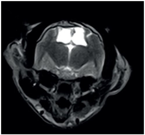
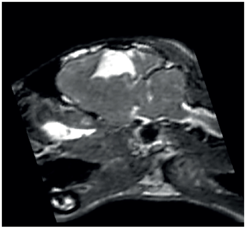
Two focal cerebral defects filled by cerebrospinal fluid and lined by white matter were observed in bilateral cerebral lobes, creating a communication between the lateral ventricles and subarachnoid space. The right defect occupied the right parietal lobe while the left one extended from the left parietal to the occipital lobes. The right-sided defect comprised the post-cruciate and lateral sulci while the left-sided cavity involved only a large extension of the lateral sulcus. Mass effect was not verified since the defect size was similar in both hemispheres. The animal of the present study did not show any neurological signs, differing from the cats of previous reports who displayed seizure and/or ataxia. A bilateral defect, as observed in the cat of this study, was reported only in one animal with ataxia who showed fluid-filled cavities in bilateral occipital lobes. In all the other cats reported in the literature, a unilateral defect was observed.
Conclusions
This study describes the MRI aspect of bilateral porencephaly in an asymptomatic mature cat.