The Pathology Associated with an Outbreak of Marek’s Disease in Green Junglefowl (Gallus varius)
Abstract
The green junglefowl (Gallus varius) is a rare bird from Indonesia that is threatened by loss of habitat and hybridization with domestic fowl. Marek’s disease virus (MDV) is a herpes virus often associated with neoplastic transformation of lymphoid cells in avian hosts (primarily chickens).1 Over an 18-month period, 13 captive-bred green junglefowl died with evidence of atypical or neoplastic lymphoid infiltrates in multiple organs. Clinical signs observed prior to death included lymphocytosis (>100,000 cells/µl in some cases), weakness, ataxia, lameness, and pallor of the comb. Disease often progressed to severe ataxia and recumbency. Most birds were euthanatized due to the progressive neurologic disease. Birds averaged eight months of age at the time of disease/death.
Gross lesions were seen in multiple organs and included patchy, pale, tan infiltrates; organomegaly and pallor; and distinct mass formation (Table 1). Gross lesions were most commonly seen in the skeletal muscle (7/13), spleen (5/13), and kidney (5/13). Enlargement of the sciatic nerves, a classic lesion of Marek’s disease, was observed in four birds. Histopathology revealed lymphoid infiltrates in numerous sites (Table 1). Histopathologic lesions were most commonly seen in spleen (12/13), kidney (11/13), lung (11/13), skeletal muscle (11/13), peripheral nerve (10/13), and liver (10/13). Lymphoid infiltrates were in some cases interpreted to be inflammatory in nature and in other cases, were obviously neoplastic. MDV infection was confirmed via PCR amplification of the oncogenic MEQ gene from abnormal sciatic nerve and muscle collected from four birds. Transmission electron microscopy confirmed the presence of viral particles in neoplastic lymphoid cells from one bird. Several birds had other diseases concurrent with Marek’s disease including intestinal coccidiosis (6/13), intestinal nematodes (4/13), systemic bacterial infection (3/13), and aspergillosis (1/13).
Table 1
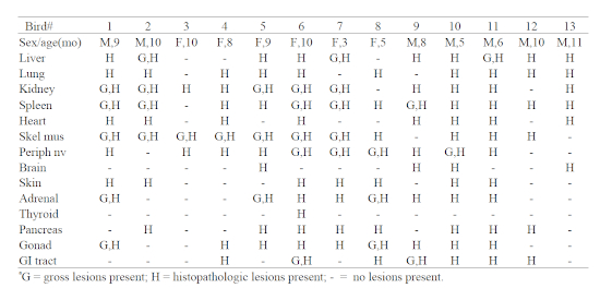
Location of pathologic lesions noted in green junglefowl (Gallus varius) infected with Marek’s disease virus
Following the diagnosis of Marek’s disease in multiple birds, a decision was made to vaccinate current juvenile and future newly hatched junglefowl. Birds were vaccinated against MDV type 3 (Fort Dodge, MD-VAC, 0.2 ml SC). The manufacturer’s recommendation is to vaccinate birds at <24 hours of age. Three <24-hour-old chicks were vaccinated, as well as four apparently healthy and serologically negative juvenile birds (two months old). Three of the four birds vaccinated as juveniles and two of the three birds vaccinated as chicks died of Marek’s disease. Since MDV was first diagnosed in this flock, 12/14 (86%) of the on-site-hatched juvenile junglefowl have died of this disease.
It is unknown whether green junglefowl are uniquely susceptible to MDV or if the MDV strain responsible for this outbreak is especially virulent. MDV has been isolated from plasma collected from affected birds, but the virus type has yet to be identified. Identification of the specific virus type may aid in future vaccination attempts for newly hatched birds. At this time, breeding of green junglefowl at this institution has been stopped until procedures to protect chicks from MDV can be developed.
Literature Cited
1. Ritchie BW. Chapter 7. Herpesviridae. In: Ritchie BW. Avian Viruses, Function and Control. Lake Worth, FL: Wingers Publishing, Inc.; 1995:200–204.