Corneal Lipidosis, Pacman Frog (Photograph)
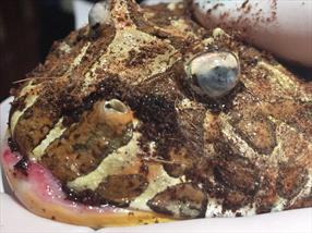
A Pacman frog with corneal lipidosis. Courtesy of Dr. Melissa Andritz.
Amphibian corneal lipidosis is a syndrome involving fat deposits on the eye; they’re also called lipid keratopathy. The deposits refer to a specific sign of a lipid (fat) storage disorder: the accumulation of cholesterol deposits in the corneal tissues. The name is also used synonymously for the systemic lipid storage disorder called xanthomatosis, which involve the deposits of a lot of cholesterol in any tissues in the body. This fat storage disease often may be diagnosed at first as an eye condition. However, the fat storage is only one sign of a systemic (whole body) lipid storage disorder.
Early stages of the disorder are seen as some white spots (stippling) on the surface of the cornea, which covers the front of the eye ball. The stippling is most commonly noticed as a faint haze or a white spot or line on one or both eyes. This haze or bits of white may progress to a blob of white material with an irregular surface that gradually covers the entire cornea.
With more advanced disease, the white blob may appear three-dimensional as a bump with a white rough surface. As lipid deposits accumulate and the bumps get larger, bleeding may occur centrally or along the edges of the bumps. Occasionally, cholesterol masses called xanthomas may be in the skin or may be detected on internal organs after shining a light through the skin.
The exact reasons for how this disorder comes about is unknown but most of the theories center on the diet fed to captive amphibians. Most amphibians are adapted to feeding on prey items that have low cholesterol levels. Most of the captive prey species fed to captive amphibians (e.g., crickets, rodents, mealworms) contain higher amounts of cholesterol than wild prey and may have a different balance of fatty acids. Amphibians may not be equipped to assimilate and eliminate these unnatural levels of lipids, and so, over time, cholesterol may accumulate in tissues other than the fat bodies—the normal storage organs for excess fat in amphibians.
Even when captive amphibians are fed prey low in cholesterols and fat, many owners overfeed their animals and many are obese. This overfeeding is also thought to be a trigger for the development of corneal lipidosis.
Husbandry problems may also play a role in developing corneal lipidosis and it is thought that captive amphibians, that are unable to attain high enough body temperatures may be prone to the disorder. Along with this, amphibians that are not reproducing do not have sufficient turnover of fat stores through egg production or through mate-attracting behaviors, and this may promote corneal lipidosis as well.
A short summary of risk factors for captive amphibians to develop cornel lipidosis and xanthomatosis include:
- Cholesterol-rich diets, such as crickets that are fed dry dog food or other cholesterol-rich kibbles or mashes
- Overfeeding, particularly of amphibians that consume rodents, goldfish, and other whole-body vertebrate food items leading to obesity
- Living in temperatures below the preferred zone
Affected Reptiles
Cuban tree frogs (Osteopilus septentrionalis) and White’s tree frogs (Litoria caerulea) are commonly affected, but any adult amphibian may develop this disorder. Older, female frogs of these species seem to get the disorder more than younger animals and males. However, many species of frogs and toads have been diagnosed with this disorder and it is likely that any captive amphibian can get this disease.
Diagnosis
Your veterinarian will start with a detailed history, husbandry review and physical examination. Often the white deposits on one or both eyes and the diet history will suggest the disorder. Your veterinarian may want to collect a blood sample to look for some chemistry values. These values include a high plasma cholesterol and elevated triglycerides.
Treatment
Your veterinarian will discuss and suggest specific treatments for your situation. The general goals and objectives are to:
- Restrict calories
- Prevent intake of excess fat
- Increase use of stored calories
- Control pain and inflammation.
Diet frequency and amounts should be adjusted so that additional fat is not accumulated. As an example, for an adult White’s treefrog, four to six adult crickets provide more than enough calories for one week. Rodents and goldfish should be eliminated from the diet. Domestic prey insects fed to amphibians should be maintained on low-fat diets such as vegetables and whole grain products, and the prey should not be allowed to consume high-cholesterol diets such as kibbled dog food.
Any husbandry deficiencies should be corrected; special attention should be placed on providing basking spots that meet or exceed the high end of their preferred temperature zone for the species, so that the amphibian can reach higher body temperatures, which may help to mobilize lipid deposits.
In some cases, your veterinarian may prescribe a pain medication such as a nonsteroidal antiinflammatory drug (NSAID) such as meloxicam as the cholesterol deposits may be painful or cause inflammation in the tissues where they are deposited.
Prevention
In collaboration with veterinarian, the best way to prevent this disorder is to provide good husbandry to your captive amphibians. Especially important is to provide an appropriate diet with good vitamin supplementation. Cultivate unique invertebrate prey sources, such as grasshoppers, springtails, and firebrats (silverfish), to offer a more varied assortment of prey than is provided by fruit flies, crickets, and mealworms.
Just as important is to provide a proper temperature gradient and thermal environment and a varied habitat that encourages natural behaviors such as mate calling and reproduction, which require additional energy to turn over stored lipids. Remember that amphibians are efficient at converting food to fat so adherence to a diet that meets but does not exceed an animal’s caloric needs is important. Even though it is fun to watch amphibians eat, it is not healthy for them to overeat!
Prognosis
Unfortunately, once an amphibian has signs of corneal lipidosis the prognosis for a complete cure is guarded to poor. Frogs that have had appropriate nutritional management and have benefited from correction of poor husbandry may live up to four years beyond detection of initial corneal opacities. Frogs that have more advanced lesions or internal xanthomas at the time of detection may not live six months.
You should have regular appointments with your veterinarian to assess the overall health of your amphibian and to monitor response to treatments. For those amphibians showing signs of chronic pain that is not responsive to NSAIDs or other forms of pain medications, discuss with your veterinarian what time euthanasia would be appropriate.