Most diabetic dogs will develop cataracts and go blind. This FAQ is designed to assist the owners of diabetic dogs in knowing what to expect and to make decisions regarding cataract surgery.
What Is a Cataract?
A cataract is an opacity in the lens of the eye. The entire lens may be involved or just a part of it. The patient will not be able to see through the opacity.
Why Do Diabetic Dogs Get Cataracts?
The lens of the eye is round, hard, and normally as clear as glass. Looking at the lens, it is hard to believe it is a piece of living tissue. The lens is suspended by fibers that can adjust its position so that one can focus vision. The lens is encased in a capsule and depends on the fluids of the eye for nutrients. The lens does not receive a direct blood supply.
Normally, the lens absorbs glucose from the eye fluids, using most of this for its own energy needs. Some of the excess is converted to another sugar called sorbitol. When there is excess sugar in the eye fluids, as in diabetes mellitus, there is excess sorbitol produced. Sorbitol pulls water into the lens, which in turn disrupts lens clarity and causes the cataract. Fructose is also produced from the excess glucose and also contributes to this water absorption.
Cataracts do not necessarily imply poor diabetic control. Even well-controlled dogs can still get cataracts.
How Long Does It Take To Go Blind?
Generally, the cataract has matured, and the dog is blind in a matter of weeks.
Until recently, blindness in a diabetic dog was basically a foregone conclusion, but there is a new product called Kinostat® that has changed that outlook. To review, the lens absorbs glucose from the fluids of the eye and uses this glucose as nutrition. Any extra glucose that is absorbed into the lens is converted to sorbitol by an enzyme called aldose reductase. Sorbitol pulls water into the lens to prevent the lens from becoming dehydrated. This is all well and good, but in the diabetic state, there is lots of excess glucose, and the excess glucose gets converted to excess sorbitol, which, in turn, pulls so much water into the lens that clarity and function are disrupted, and a cataract is formed. Kinostat is an aldose reductase inhibitor that curtails sorbitol production. Early use of Kinostat may significantly delay or even completely prevent the development of cataracts.
Kinostat is preventive only and will not reverse cataract formation that has already occurred. Kinostat is unfortunately not yet commercially available, though has been reportedly near coming to market for some time.
What Does It Mean To Say That a Cataract Is Mature?

A mature cataract. Image Courtesy of Dr. Rhea Morgan.
A cataract’s maturity is determined by how much visual impairment the pet is believed to have. Since we cannot ask a dog to read an eye chart, we must determine this by visually inspecting the eye. A light is used to look into the eye and view the colorful area at the back of the eye called the tapetum. (This is the area that flashes or appears colored in certain lighting.) When less than 10 percent of the tapetum is obstructed, the cataract is very young and does not significantly change vision. When 10-50 percent of the tapetum is obstructed, this cataract is called early immature. When 51-99 percent is obstructed the cataract is late immature. The mature cataract obstructs the entire tapetum. Ideally, a cataract is removed in the early immature stage for the lowest surgical complication rate.

A mature cataract. Image Courtesy of Dr. Jane Bondt
When a cataract is hypermature, it starts to liquefy and dissolve. This can actually lead to restoring vision, which sounds like a positive turn of events; unfortunately, the dissolution process is quite inflammatory. The resulting inflammation can be painful and can damage the eye even more.
All cataracts do not progress all the way to hypermature and may stay static or progress at changing rates; however, diabetic cataracts are notorious for reaching hypermaturity and creating inflammation.
What Is Uveitis?
Uveitis is inflammation of the eye's uveal tract, which consists of the eye's vascular tissues. In this situation, uveitis is the inflammation that results when the hypermature cataract begins to liquefy. Uveitis is painful and tends to cause the eye to become reddened and the pupil to constrict. When a pet has uveitis before surgery, success (pain-free vision) occurs in only 50 percent of cases 6 months after surgery as opposed to 95 percent of cases for whom there was no uveitis pre-operatively.
Can My Dog's Vision Be Restored?
Maybe. It depends on the health of the eye, excluding the cataract. If the only thing wrong with the eye is the cataract and the patient's diabetes mellitus is well-regulated, surgery can remove the cataract and place an artificial lens. If the eye is deeply inflamed or is otherwise damaged, the eye may be blind even without a cataract. A veterinary ophthalmologist can evaluate the eye to determine the feasibility of cataract surgery.
Eye Phacoemulsification
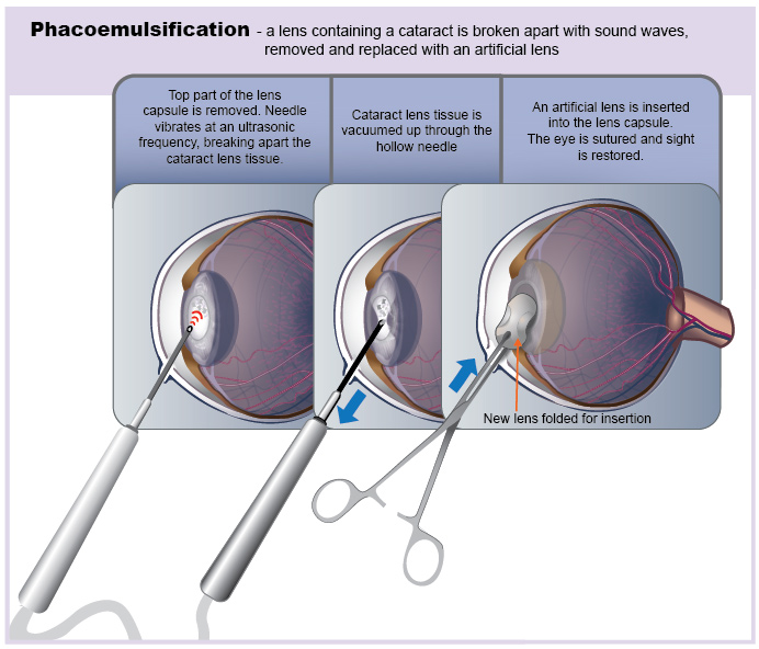
Illustration by Tamara Rees of VIN.
It is important to note that there are numerous eye drops advertised that claim to dissolve cataracts. None of these products actually work and wasting time with them may actually allow uveitis to set in and make for a much worse prognosis for vision.
Is it Cruel To Keep a Dog Blind?
Not at all. Dogs do not depend on vision the way humans do. A blind dog can get along very well as long as the furniture isn’t moved and the dog is properly supervised.
For tips on helping a blind dog adapt:
Blind Dog Support
There are many medical conditions that render a dog blind and as long as the condition is not painful, the dog can live a normal life as a successful and happy pet.
Cataract Surgery: What Is The First Step?
The first step is a consultation with your regular veterinarian. Your dog’s diabetes must be well-regulated before surgery is considered. If pre-operative lab tests show nothing to preclude anesthesia, your veterinarian may refer you to a veterinary ophthalmologist, as clinics do not usually have the specialized equipment necessary. A regular veterinarian is not qualified to perform cataract surgery.
What Happens At The Ophthalmologist's?
It is necessary to determine if the eye is going to be visual after cataract surgery. There is, after all, no point in performing this surgery if the eye is going to be blind anyway. The most important test is called an ERG (an electroretinogram). This test checks the retina for electrical activity, which indicates the eye should be able to see after the cataract is removed.
In addition to the ERG, the ophthalmologist will check for uveitis. It should be treated before surgery to minimize the inevitable inflammation after surgery.
What Kind of Surgical Procedures Are Done?
There are two types of surgery: lens extraction and phacoemulsification. With lens extraction, the incision tends to be larger, the post-operative inflammation is greater, and the potential for leaving bits of the lens behind is also greater.
With phacoemulsification, an ultrasonic instrument is used to liquefy the lens, and a sort of vacuum cleaner sucks the lens away. This procedure is more difficult if the patient is older, and the lens is thus harder in consistency. This method is preferred for diabetic patients.
After either surgery, an artificial lens is usually placed for optimal post-operative vision.
What Kind of After Care Is Needed?
The patient will need to wear an Elizabethan collar after surgery to protect the eye. Cortisone eye drops are needed for probably several weeks. Oral anti-inflammatories will be needed for weeks to months after. Drops to keep the pupil dilated will also be used.
What Kind of Complications Are Possible?
Complications to consider are:
- Long-term uveitis (probably of most concern for diabetic patients)
- Opacification of the lens capsule (usually correctable with a laser)
- Corneal clouding (can be managed with 5 percent saline eye drops 4 to 6 times daily)
- Bleeding in the eye
- Glaucoma
- Retinal detachment (particularly if the cataract is hypermature)
Should Surgery Be Done On Both Eyes?
It is important to remember the old saying that the one-eyed man is king among the blind. A dog need only have one cataract removed to have vision restored. Doing both eyes is an option to discuss with the ophthalmologist, as some dogs need all the vision they can get.
Cataract surgery requires committed patient care both in the hospital and at home. Your veterinarian will be able to answer your questions or direct you to other appropriate resources.