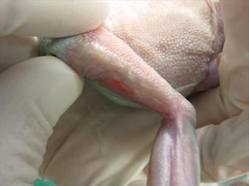
Frog with discoloration and wound of the skin due to worm infestation. Photo courtesy of Dr. Richard Levine.
Amphibian nematodiasis is caused by infection with parasitic worms called nematodes. Infections may occur in any tissue, organ or body cavity. Nematodes infecting the intestine and lungs cause the worst disease in captive amphibians. Infections with nematodes are commonly known by various other names including: helminthiasis, lungworms, Rhabdias infection, Strongyloides infection, hookworms, roundworms, and filarid worms.
Nematodes have a variety of life cycles that are important for diagnosis, treatment, prognosis and prevention. The two common types of life cycles are called direct and indirect. A direct life cycle means that the nematode can infect the amphibian without any help, while a nematode with an Indirect life cycle needs another species, such as an insect or another animal, to be able to become infective to the amphibian. For captive amphibians, the nematodes with direct life cycles are the ones that cause disease most commonly, and they often cause bad infections called superinfections. The most common direct life cycle nematodes in captive amphibians are Rhabdias and Strongyloides.
Rhabdias and Strongyloides are the important disease-causing nematodes in in captive amphibians owing to their direct life cycle and the fact that they are self-fertilizing (can have young on their own without another nematode) and are thus able to cause superinfections. Rhabdias is a lungworm while Strongyloides infect the intestine. Direct damage to various organs by migrating larvae and adult worms may result in secondary bacterial infections and debilitation. Low levels of worms may be harbored without clinical signs of disease. Because this class has free-living generations that can survive in the soil, reinfection can occur from plants and other organic material in the enclosure. Superinfection results from stress, especially overcrowded enclosures.
It sounds bad enough for your amphibian to have worm infestations in the lungs, skin, or eyes, but if they could infect you as well that would be a whole different story. Fortunately, amphibian nematodes are not known to be zoonotic; they can’t infect you.
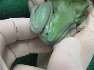
Frog with open wound or ulcer due to worm infestation. Photo courtesy of Dr. Richard Levine
How can you tell if your amphibian has nematodes? This is not a straight forward answer but certain signs are a clue. These signs include: weight loss and looking skinny; sudden death, which is common with intestinal and lung infections; diarrhea; bloating; buoyancy problems (especially in tadpoles- they may float lopsided or upside-down); and skin infections.
Your amphibian may go from having bright coloration to a dull coloration. If the worms are infecting the mouth, you can see salivation or slobber. On the skin, you may see white vesicles or blisters; sloughing of skin; and open wounds or ulcers. You may notice worms in the eye or moving under the skin. Commonly, there will be small nodules in the skin or on organs. These are known as granulomas and nematodes are inside them.
Affected Reptiles
All captive amphibians can get nematodiasis. Common risk factors that lead to disease are stressed and debilitated amphibians; lack of quarantine and exposure of collection amphibians to new, wild-caught amphibians; and high stocking densities in the enclosures.
Diagnosis
To diagnose nematodiasis, your veterinarian will begin with a thorough husbandry history and physical examination. Your veterinarian will also do some testing to detect any nematodes. Common tests performed in the clinic include: examination of the feces, mucus from the mouth, material from the body cavity, and skin scrapings to look for worms or their eggs.
Commonly, your veterinarian will use a small light source to shine through the amphibian, which is called transillumination, to detect nodules (granulomas) on the organs inside the body. In some cases, your veterinarian may want to collect blood and take a set of x-rays. As mentioned above, the physical exam findings are often non-specific but if your amphibian has lumps, bumps or non-healing ulcers, that will help make nematodiasis a possibility.
Additional tests might include a biopsy of one of the lumps to look for nematodes in the tissue.
Treatment
Once an infection with nematodes is confirmed, specific treatments can be initiated. The goal of treatment is to make the amphibian feel better by reducing the numbers of adult nematodes as it is impossible, in most cases, to eliminate all nematodes. Use antibiotics to eliminate any bacterial infections and any disease with other intestinal parasites, such as amoeba and coccidia. Provide nutrition if the amphibian needs it.
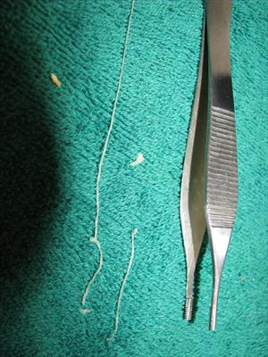
Worms removed from the frogs shown above. Photo courtesy of Dr. Richard Levine
Your veterinarian will decide which drug is best to use for the nematode infection and species of amphibian. The drugs used to treat nematodes are call anthelmintics and include ivermectin, fenbendazole, levamisole and milbemycin. Depending on the drug selected, it may be given topically on the skin or orally.
If your amphibian is dehydrated, your veterinarian may bathe them in a solution to help rehydrate them and, if needed, nutrition can be provided via tube feeding.
Because nematodes can spread from one amphibian to another, your veterinarian may suggest treating all the amphibians in a particular cage or group to prevent transmission of the disease to the healthier amphibians.
As always, your veterinarian will discuss any improvements needed to husbandry with particular attention to sanitation and will suggest that all the substrate and cage props be changed out for new, clean material.
Prognosis and Prevention
The prognosis for amphibians with nematodiasis is always guarded because many nematodes are resistant to anthelmintic drugs and may be reduced in number but not eliminated, and so re-infections are common and need to be monitored for on a regular basis.
All incoming amphibians should be considered sources of nematode infection. With your veterinarian, develop a quarantine program to detect carriers before they are introduced to a trouble-free collection and preventively give anthelmintics to all incoming amphibians.
As always, husbandry should be of the highest quality. Some amphibians may become stressed from too frequent intrusion into their enclosures. Maintaining balance between appropriate sanitation (i.e., removing fresh fecal droppings from the cage) and a low-stress environment is difficult. Review specific husbandry needs for the species you have or are going to acquire. It’s really important to talk to your veterinarian about husbandry and to research husbandry needs of the particular species you have.