Echocardiogram of dog's mitral valve
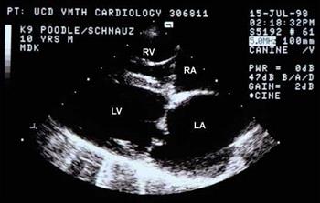
Photo courtesy of Dr. Mark Kittleson
What is an echocardiogram?
An echocardiogram, also known as an echo or cardiac ultrasound, is a diagnostic tool that looks closely at the heart as well as inside and around it. An echo uses high-frequency sound waves to create live images, allowing veterinarians to get an idea of what the heart looks like and how it is functioning in real time. This provides information about the size, shape, and function of the heart, its four chambers, the heart valves, and surrounding structures, such as the pericardial sac.
Doppler (both Color Doppler and Spectral Doppler), is another non-invasive ultrasound test used to assess how blood is flowing through the heart, as well as how blood enters and exits it.
Can my regular veterinarian do an echocardiogram?
An echo is a little bit different from a general ultrasound because it requires a great deal of knowledge and training to perform, is more technically difficult, and sometimes requires specialized equipment such as cardiac transducers (ultrasound probes). This form of imaging is tightly focused on the heart. Other organs, such as those in the belly, are rarely examined during an echocardiogram.
In addition to assessing for obvious abnormalities (e.g. masses in and around the heart, fluid in the pericardial sac), measurements of individual heart wall thickness, chamber size, and blood flow are taken. Calculations are done to see how well the heart is functioning based on these measurements. As such, many general practice veterinarians will refer pets to veterinary cardiologists or other imaging specialists for echocardiograms rather than perform the procedure themselves.
What is the procedure?
Echocardiograms are typically done with the pet lying on an ultrasound-specific table. The ultrasound transducer (probe) is held against the skin overlying the heart. The transducer sends sound waves to the heart, which are reflected back to the transducer and translated to images on a screen. Hair does not conduct sound waves very well, so the pet’s skin is usually moistened with alcohol prior to the procedure. Ultrasound gel is then applied to the skin to provide better conduction. Ribs do not conduct sound waves well either, so the transducer is often placed in many strategic areas on the skin between the ribs to get an accurate view of the entire heart.
Echocardiograms are typically painless and often done in a quiet, dark room. Most pets are able to lie comfortably without stress and with minimal restraint. Rarely do pets need sedation. Almost all pets can safely undergo echo, but your veterinarian will be the best judge.
Why would my pet need this procedure?
If your pet was recently diagnosed with a heart murmur or is suspected to have heart disease, your veterinarian may recommend an echocardiogram. This recommendation will be based on factors such as concerns seen on X-rays and associated clinical signs like coughing, shortness of breath, or fainting.
A heart murmur in a young, healthy animal usually warrants an echocardiogram at some point to determine what congenital disease (if any) is causing the murmur, and how severe the disease is. This is important because certain congenital diseases, which the animal is born with, can be repaired or improved with various interventions.
Physiological or innocent murmurs are harmless murmurs that are created from blood flow patterns through the heart chambers. They can sometimes be heard unexpectedly, especially in young puppies and kittens, and are not a cause for concern. However, physiologic murmurs are not easy to differentiate from heart murmurs associated with heart disease or dysfunction. Without any clinical signs, your veterinarian may recommend monitoring the murmur until the next vaccine series or checkup. If your veterinarian continues to hear a murmur by eight weeks of age, even if your dog or cat is showing no signs of heart disease, an echo will likely be necessary. Some veterinarians may recommend waiting until your pet is a bit older, up to 16 weeks of age, to allow for an adequate echocardiogram without requiring a lot of handling and stress to the pet, but usually only if your pet remains otherwise healthy and without any signs of heart disease.
The echo can show if the heart is working properly, and if not, what the problem is. Medications and treatments for heart disease are tailored to the individual, so understanding the problem correctly helps provide the best treatment. Echocardiograms can also be used to see if treatments are helping or if a change in dosage or new medications are required.