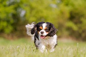
Cavalier Spaniel
(Suh-ringo-my-eelia)
Also called caudal occipital malformation syndrome, COMS, Chiari-like malformation, occipital dysplasia
Syringomyelia is an odd word, and it does not exactly roll off the tongue on the first attempt. Furthermore, most of us never need to know what it means. Who needs to know what it means aside from doctors and those with a special interest in neurology? Probably anyone contemplating owning a Cavalier King Charles Spaniel should know what it means since it appears a large percentage of this breed has the condition. This condition is not limited to the Cavalier King Charles Spaniel, though numerous individuals of other breeds have been affected. Syringomyelia causes pain in the back of the neck as its hallmark sign but can also produce other neurologic deficits and weaknesses. It is a progressive condition.
Neurology is a complicated science, but fortunately, one can understand this condition without specialized knowledge. Basically, syringomyelia involves small fluid pockets within the spinal cord and is caused by a mismatch between skull size and brain size. To understand how this leads to pain in the back of the neck, we'll review a little basic anatomy.
The Central Nervous System
The nervous system is divided into two parts: the central nervous system (brain and spinal cord) and the peripheral nervous system (nerves). The brain is located in a cavity in the skull where it is well-protected and bathed in cerebrospinal fluid. The spinal cord runs down a canal formed by our vertebrae (our backbones). It, too, is bathed in cerebrospinal fluid.
The cerebrospinal fluid, lovingly referred to as CSF, provides nutrition, waste removal, lubrication, and shock insulation to our central nervous system. You might consider that the brain and spinal cord are almost like fish in an aquarium, and CSF is the water.
CSF is formed in chambers inside the brain called ventricles. These chambers generate CSF, which then flows down a central channel (called the central canal) down the middle of the spinal cord, then outside and around the spinal cord and brain, and is eventually absorbed into the bloodstream. The blood-brain barrier separates the materials in the bloodstream so they are not secreted into the CSF. This is protective but also keeps many potentially helpful medicines out of the central nervous system.
So, What is Syringomyelia?
In syringomyelia, there is altered CSF flow due to an assortment of problems (congenital malformation, tumor, trauma, etc.). The altered flow leads to cavities of fluid forming in the spinal cord. This creates swelling and, ultimately, small pockets of fluid within the spinal cord, and since the cord is confined within a bony protective “cage,” there is no room for the swelling to expand. Pain results.
Why is this Sometimes Called the Chiari Malformation?
The Chiari malformation is by far the most common cause of syringomyelia, and because the two conditions frequently go together, it is not unusual to see the terms used interchangeably, though this is not technically correct.
Dr. Hans von Chiari was a pathologist who, in 1891, categorized different congenital malformations in human infants that generated the condition we now call syringomyelia. When human medical terms are applied to veterinary patients where anatomy is similar yet different, it is questionable whether the same terms are really appropriate. This is another reason why the trend in veterinary patients is towards the term Chiari-like malformation and away from Chiari malformation.
A dog can have the Chiari malformation without syringomyelia. The Chiari malformation is the most common cause of syringomyelia but not the only cause.
What is Happening in Syringomyelia?
The driving force for creating the abnormal fluid pocket seems to be the beating of the heart and the pulse it generates. Each pulse generates a pressure wave in the CSF, displacing fluid from the brain and down into the spinal cord. This is how CSF normally accomplishes circulation, but in syringomyelia patients, there is some kind of obstruction to CSF flow (usually the Chiari-like malformation). This creates increased CSF pressure around the obstruction, and CSF can be pumped directly into the spinal cord tissue. This distends the cord, creating fluid pockets. The fluid is not CSF but simply what is called extracellular fluid. The distended cord exacerbates the obstruction of CSF flow, thus creating a progressive problem (i.e., a vicious cycle).
Why is this a Back of the Neck Problem in Cavalier King Charles Spaniels?
The brain is commonly thought of as one organ, just as the skull is often thought of as one bone. In fact, the brain has many parts, all very different, and the skull consists of many bones fused together. The occipital bone is the skull bone in the back of the head. It contains a depression known as the caudal fossa in which the cerebellum, pons, and medulla all take up space.
In Cavalier King Charles Spaniels, the common cause of syringomyelia is an abnormal shape of the caudal fossa. Basically, the cerebellum is too big, and the caudal fossa is too small. This compresses the cerebellum, pons, and medulla, creating the obstruction that allows for syringomyelia to occur. In this case, the fluid pocket is just at the back of the neck. The abnormally shaped caudal fossa is the Chiari malformation.
What Does Syringomyelia Mean for the Patient?
In short, the fluid pocket in the spinal cord hurts. Certain postures, states of excitement or even weather conditions can make the situation worse or better. It would seem that the pain would be symmetrical behind the neck, but it is not. Scratching at the neck, chest, or shoulder without actually making contact with the skin and doing so only on one side is the most commonly reported observation. People with syringomyelia report headaches, neck pain, back pain, facial pain, or pain radiating down an extremity. The neck can develop a curve away from the lesion.
Aside from pain, there may be other signs of spinal disease, such as weakness in the legs. Facial nerve paralysis (leading to loss of facial expression) is common in the Cavalier King Charles Spaniel, with or without syringomyelia. Recent technological advances that made magnetic resonance imaging (MRI) more accessible to veterinary patients have shown that syringomyelia is not an uncommon problem in this breed, and it has been suggested that there may be a connection with facial nerve paralysis previously thought to be unexplainable. Seizure disorders are also common with this breed, so we do not know if there is an association with syringomyelia or if this is a coincidence.
Most patients with caudal fossa overcrowding are diagnosed between the ages of six months and three years.
Confirming Diagnosis/Ruling Out PSOM
Diagnosis cannot be made without magnetic resonance imaging (MRI). This not only confirms that there is a fluid pocket but also helps determine the cause of the syringomyelia.
There are other conditions that can cause neck pain: disk disease, granulomatous meningoencephalitis (GME), disk infection, tumors, vertebral instability, and even ear or skin disease. One particular condition that might mimic syringomyelia is called primary secretory otitis media, or PSOM, and the Cavalier King Charles Spaniel is genetically predisposed to this condition, just as it is to syringomyelia.
PSOM also causes neck scratching and/or facial nerve paralysis as well as head shaking, hearing loss, head tilt, or any combination of the above. PSOM is a condition that leads to the accumulation of thick mucous discharge in the middle ear and currently, the only treatment is periodic lancing of the eardrum and flushing out the mucus. The leading theory is that the conformation of the Cavalier's throat leads to abnormal fluid drainage through the auditory tube connecting the ear and throat. Obviously, it is important to distinguish syringomyelia from PSOM. If the dog has an obvious bulging ear drum (generally easily visible through a routine otoscope), this largely confirms PSOM; however, a good many dogs with PSOM do not have an obvious bulging ear drum. For these dogs, MRI should be able to distinguish these two conditions.
Treatment: Surgery
Probably the most effective way to address syringomyelia is through surgery to decompress the CSF flow obstruction. The earlier surgery is performed the better the results. The largest study to date looked at 16 dogs receiving surgery, and 81 percent experienced success. The problem is that scarring tends to lead to recurrence, and of these 16 dogs, only 45 percent were still judged as having good life quality two years after surgery. The fluid pockets eventually reform. Pain management medications, as described below, are almost always needed despite surgery. Simply draining the fluid pockets has not been meaningfully successful.
Treatment: Drugs

Image Courtesy of MarVistaVet
Obviously, surgery is invasive and expensive and may not create long-term results in a given patient. Given this information, many opt for medical management of syringomyelia, which means pain relief and using medication to reduce CSF pressure.
There are three approaches to using medication: addressing pain, reducing CSF formation, and reducing swelling with corticosteroid hormones. Because of interactions and side effects, it is likely not practical to try all three approaches simultaneously. You can begin with a medication such as furosemide and/or omeprazole to reduce CSF production. Add a pain reliever such as gabapentin, which addresses neuropathic pain specifically, and possibly an anti-inflammatory pain reliever (NSAIDs). Dosing can be adjusted with the diuretic several times before deciding if it helps or not. Corticosteroids can be added in, assuming the other medications are compatible with them, so as to find an effective regimen.
Additionally, food bowls can be elevated, and the patient's collar can be changed to a harness to avoid pressure on the neck.
Despite these efforts, 75 percent of patients will continue to deteriorate if surgery is not included in the treatment plan.
If a combination of pain medication and diuretics is not helping, the pain medication can be replaced by a veterinary non-steroidal anti-inflammatory (NSAID). There are many options, and your veterinarian can guide you through the choices.
Corticosteroids such as prednisone can be used with gabapentin but not with the NSAIDs. It is unclear exactly what the steroids do in this situation: reduce CSF production, reduce inflammation, simply blunt pain, or something else, but they are an additional combination that can be used. Long-term use of steroid hormones has important side effects to consider.
Omeprazole, mentioned previously, has been helpful in treating syringomyelia, but its use is best restricted to 8 weeks in duration.
Omeprazole is a strong stomach antacid that is also able to reduce CSF secretion. If the stomach does not require such a powerful antacid, it may be harmful to use one long-term in this way, but the spinal pain may be helped.
Patients without Pain
There are three reasons to screen an apparently normal Cavalier with an MRI: evaluation as a candidate for breeding, general evaluation, and monitoring of the individual. Whether or not you need to monitor an apparently normal individual is a controversial matter as MRI requires anesthesia and is, therefore, not entirely without risk. Dogs that are not experiencing pain do not need treatment.
As for breeding, it is important not to breed affected individuals whether they are experiencing pain or not. This is problematic as some dogs do not develop their condition until later in life after they have already been bred. Still, the U.K. Cavalier clubs have developed a screening protocol involving screening MRIs. The U.S. clubs have thus far not adopted these guidelines but that does not mean they cannot be followed or adapted.
Our understanding of this condition is still incomplete, and research is still active. For other Internet-based support, see:
Cavalier Health
The Cavalier Health Club (UK)
Cavalier Health Foundation