Dalen W. Agnew, DVM, PhD, DACVP
Department of Pathology, Microbiology, and Immunology, University of California, Davis, CA, USA
Introduction
Examination of the central nervous system, both grossly and histologically, is an important component of a complete necropsy. Cerebral nematodiasis, West Nile virus infection, rabies, distemper, and organophosphate toxicity are just a few of the possible diseases with serious herd and public health significance, which may only be diagnosed by careful analysis of the brain and/or spinal cord. Removal of the brain is strongly suggested for a complete necropsy, and though it may appear a daunting task, a few guidelines and power tools will allow efficient removal of the brain and a complete necropsy.
It is usually preferred that the brain be removed whole by removal of the skull cap. This technique has been well documented in necropsy texts and is commonly taught in veterinary schools.2 Briefly, after skinning the skull, a saw or ax may be used to cut on either side from the foramen magnum and the occipital condyles cranially and dorsally in a circular pattern (Figure 1). This technique is useful to examine the brain in situ and remove it whole, but unfortunately requires skinning of the head, can be time consuming, and is almost impossible to complete in rhinoceros and elephants. There are many alternative approaches to brain removal, but the author has found the following methods using commonly available tools are quick, leave a relatively intact skull, and the brain itself is removed in two parts. Certainly, the techniques presented here can be adapted to the individual preferences of the prosector and to other similar species. If nothing else, a discussion of brain removal techniques will reinforce the importance of collecting a complete set of tissues during a postmortem examination.
Figure 1. Equid skull
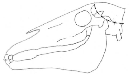
To remove the brain whole, it is necessary to cut the skull cap from the occipital condyles cranially and dorsally to the frontal bone on both sides, remove the cap, and dissect out the brain.
Tools
Some recommended tools:
1. A scalpel or small necropsy knife
2. A large buck or bow saw (ideally, with a blade designed to cut bone)
3. A carpenter’s electric reciprocating saw (Milwaukee Sawzall® or similar, with a supply of 9” wood/nail-cutting blades)
4. A Jarvis Wellsaw
5. A generator to use power tools in remote locations and field conditions
6. A hand keyhole or jigsaw can be used in the field
7. An ax or hatchet can also be used, but the brain may be damaged and the physical effort required is much greater
8. A thin, long metal spatula for removing the brain from the brain case
Equids
After removing the tongue, trachea, and esophagus (the “pluck”) from the oral cavity and neck region, the neck should be over-extended, exposing the spinal cord ventrally through the atlanto-occipital joint and the cord severed by a stab incision with a knife or scalpel. The skin on the head is cut in a line parallel to the caudal margin of the mandibular ramus through a point 1 cm cranial to the ear canal (see Figure 2, line marked A). Then using a saw, the head is cut transversely all the way through. The calvarium is now open, and the brain is in two pieces. Using a thin metal spatula, a knife, or gloved finger, the brain can be separated from its attachments in the skull and pulled free. The bony tentorium may need to be broken down with a knife to remove the cerebellum. If the cut is accurately made, the pituitary gland should be immediately adjacent to the cut in the cranial half.
Figure 2. Diagram of the intact head and skull of an equid (left) and bovid (right)
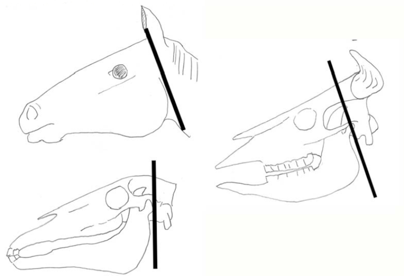
A single saw cut through the skull parallel and just caudal to the margin of the ramus of the mandible (immediately cranial to the ear canal) will expose the brain. After breaking down the bony tentorium separating the cerebrum and cerebellum and cutting the spinal cord with a stab incision through the ventral atlanto-occipital joint, the brain can be manually removed.
Artiodactylids
A similar technique as that described for equids can be used in many artiodactylids (Figure 2). This is particularly useful when antlers or horns make removal of the skull cap impossible. The precise division of the brain may vary in different species.
Rhinoceros
Rhinoceros brain removal can be challenging, particularly since many institutions want to display the skull after the necropsy The following technique (Figure 3) was successfully used to remove the brain of a white rhino, but would likely work for other species of rhino.
The skin should be removed from the dorsum of the head. Make two transverse cuts in the skull as diagramed (Figure 3, A). The first cut should be in a transverse plane drawn through the ear canal. The second cut is made in a parallel plane 14 cm cranial to the first and approximately 15 cm caudal to the eye. These cuts are in an arc so as to minimally damage the brain and extend laterally to a point 4 cm ventral to the longitudinal plane through the ear. Two lateral cuts in the longitudinal plane then connect the ends of the two transverse cuts. It is easiest to start these cuts with the Stryker saw to get a groove in which to place the blade of a larger saw. Before the brain can be removed from this access, the spinal cord must be severed at the atlanto-occipital joint, either by removing the head or with a knife through the ventral dura at the joint.
Figure 3. Head and skull of a white rhino
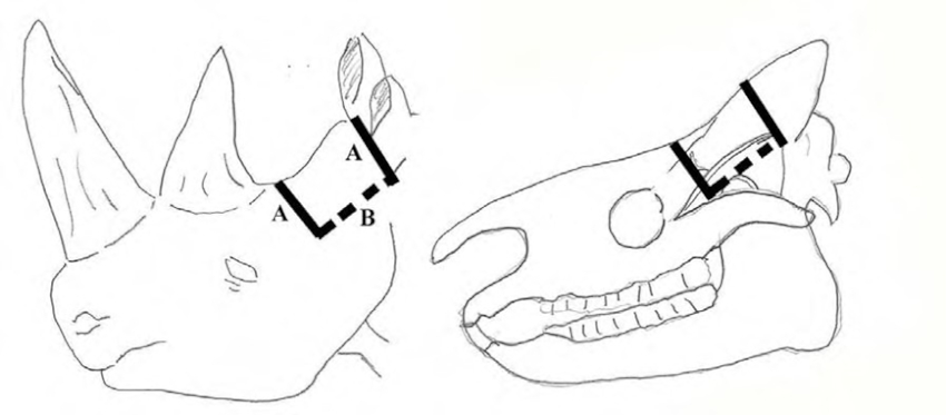
Using a reciprocating saw, cuts are made transversely (A) through the ear canal and through a point 14 cm cranial (approximately halfway between the eye and the ear). The ventral extension of these cuts can then be connected (B) on either side and the cap removed, exposing the brain below.
Elephant
Due to their massive size and the remarkable bony sinuses overlying the elephant skull, the brain is especially difficult to remove. However, the following procedure has produced good results and can be performed with either power tools or hand tools in the field.
The elephant should be in lateral recumbency to best proceed with brain removal.1 Remove all the skin from the neck to the eyes and over the top of the head. Cut down to the atlanto-occipital junction from the ventrum after removing the pluck. Once the atlanto-occipital joint is found, the head can be rolled forward slightly to open the gap between the atlas and the occipital condyles. Drive a sharp knife into this gap to sever the spinal cord to prevent further histologic stretch artifacts in the brainstem and spinal cord and make the brain easier to remove. The atlanto-occipital joint can be completely cut at this point. Clean all the muscle from the caudal aspect of the skull so that only bone is exposed approximately 15 cm dorsal and 10 cm lateral to the foramen magnum. Using the saw, make the following cuts (see Figure 4):
1. From the ventral lateral aspect of the foramen magnum, cut dorsally 15 cm at a 30° angle from the ventral midline on both sides.
2. Connect the dorsal extent of both cuts with a third cut. In order to cut through the bone, it is best to lay the saw flat on the surface of the bone until it cuts through into the cranium. Then the cut can be extended laterally to connect with the first two cuts. The bone fragment can now be removed. A pry bar may be used if necessary to separate the bone if the cuts are not complete.
3. If the resulting opening is not wide enough to remove the brain, two additional cuts may be used to widen it. Cut laterally from the base of the foramen magnum approximately 8 cm. Then cut from the cranial-most aspect of the previous cuts ventrally to the end of these two lateral cuts (essentially cutting off the occipital condyles).
Figure 4. Caudal view of an elephant skull
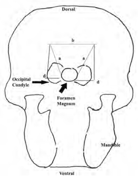
Using a reciprocating saw, cut from the base of the foramen magnum dorsally and cranially on each side for approximately 10 cm (a). Connect the dorsal aspect of both lateral cuts (b). If necessary, the remaining occipital condyles can be removed so that the brain can be more easily reached.
(Reprinted with permission from Biology, Medicine and Surgery of Elephants. Fowler and Mikota, eds. 2006).1
The cerebellum and brainstem are now exposed. Reach in with the knife or a scalpel and separate the cerebellum and brainstem from the cerebrum. The cerebellum can now be removed carefully, cutting cranial nerves as it is elevated. The cerebrum can be extracted by reaching in with a scalpel or simply a gloved hand, carefully separating the brain from its dural adhesions, and delivering it through the widened foramen magnum (much like delivering a baby). It may be necessary to separate the two hemispheres. Usually, the pituitary gland remains and can be removed by carefully lifting it by the dural attachments with forceps and using a scalpel to peel it from the underlying bone.
Brain Fixation
Like all tissues, the brain is best preserved in 10% NBF at a 10:1 ratio (formalin:tissue). Some pathologists, however, fix large brains in 40%. Unless the gross morphology is critical, it is best to separate the cerebral hemispheres and fix them and the cerebellum separately. Even though the brain may be autolyzed or accidently macerated, it should be preserved in formalin and examined. Many diagnoses can still be made from these tissues. If rabies is a differential diagnosis, the brain should be submitted fresh to the local or state laboratory. Fixation will make rabies testing impossible (although select samples may be fixed for histology when the brain is first removed).
Acknowledgments
I thank Dr. Rob Fairley for introducing me to the technique of brain removal in horses and Drs. Linda Lowenstine and Linda Munson for their advice and support during many long elephant necropsies. I also thank Drs. Murray Fowler, Susan Mikota, and Dick Montali for their kind permission to reprint diagrams from their textbook.
Literature Cited
1. Fowler M.E., and S. Mikota. 2006. Biology, Medicine and Surgery of Elephants. Blackwell Publishing, Oxford. P. 576.
2. King J.M., D.C. Dodd, L. Roth, and M.E. Newson. 2003. The Necropsy Book, 3rd ed. Charles Louis Davis, DVM Foundation, Gurnee, IL. P. 217.