
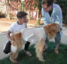
This position was the only one in which Kodee was comfortable prior to surgery. Photo by Linda Pace.
Pneumothorax is a collection of air or gas in the space surrounding the lungs. The extra air doesn’t allow the lungs to inflate normally, so dogs and cats with pneumothorax can have difficulty breathing (dyspnea), an increased respiratory rate, exercise intolerance, chest pain, cyanosis, collapsed lungs, etc. If only a tiny bit of air gets inside the chest cavity, it can be resorbed and the pet can heal without medical help. But if there is a lot of air in the chest cavity, it must be removed medically. If severe cases are left untreated, the pneumothorax can be fatal.
Pneumothorax may result from chest trauma, excessive pressure on the lungs, or underlying lung disease (asthma, chronic obstructive pulmonary disease, etc.). In some cases, the cause is never discovered.
Types of Pneumothorax
The three forms of pneumothorax are:
- A hole in the chest wall that allows air inside the open chest cavity
- The chest wall doesn’t have a hole, but there’s one in the lungs, bronchi, trachea, or esophagus
- A rip in the pleura (the membrane in the lungs) that acts like a one-way valve (lets air in, but doesn’t let it exit).
Specifically, the types can be categorized as follows:
- Closed pneumothorax: air is leaking from a hole in a lung cyst, bronchus, trachea, esophagus, or lung tissue into the chest cavity.
- Open pneumothorax: air is entering the chest cavity via an open wound in the chest wall. Causes include animal bites, gunshot wounds, puncture wounds, and automobile accidents.
- Iatrogenic pneumothorax: air entered the chest cavity during lung surgery or procedures.
- Spontaneous pneumothorax: (no known cause for the air in the chest cavity).
- Tension pneumothorax: air entered the chest cavity from the bronchus, but cannot leave the same way, thus causing progressive collapse of the lung
Surgeons removed one and a half lung lobes from Kodee, who recuperated wonderfully. Photo by Jack Pace.
Diagnosis
Diagnostic tests include patient history, physical exam (respiratory distress, coughing, difficulty breathing, traumatic wounds, etc.), chest imaging (radiography, CT, ultrasonography, MRI), and removing air from the chest cavity using a syringe (thoracentesis).
Treatment
Treatment depends upon the type of pneumothorax and how bad it is. Minor cases may resolve with 1 to 2 weeks of cage rest. Severe cases are a real emergency, and your veterinarian will probably give oxygen immediately; dogs and cats are placed in an oxygen cage. Your veterinarian may need to put in a chest tube to remove the extra air from the chest. Surgery may be needed to close up large holes in the chest, to fix injuries, or to remove damaged lung lobes (lobectomy). Unlike people, who have five lung lobes, dogs and cats have seven lobes.
Prognosis
The prognosis depends on how severe the case is and how early treatment was given.