Abstract
Radiographic evaluation of the pectoral and pelvic girdles in chelonians is often complicated by the superimposition of the shell structures. The following report describes two cases where computed tomography (CT) was used in addition to radiography.
Case 1
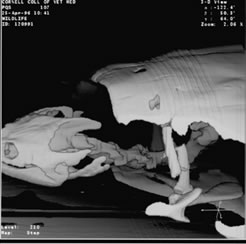
A long-term captive, 4.6-kg, adult, 11-yr-old, male, radiated tortoise (Geochelone radiata) was admitted for evaluation of a 2-wk history of shifting lameness and muscle weakness. The animal was reported to be in good body condition with decreased appetite. Initial evaluation included physical examination, hematology and plasma chemistry analysis, fecal analysis and culture, and whole body radiography. On physical and radiographic examinations, diffuse swelling of the left pelvic limb was observed. During blood analyses, hyponatremia, hypochloremia, hyperproteinemia and a mild leukocytosis were detected. During radiography, moderate-to-severe intestinal distension was detected. Therefore, contrast radiography was performed (60% w/v barium sulfate suspension PO). Transit time through the stomach and the small intestine was clinically normal. Transit time through the large intestine presumptively was delayed. This finding was attributed to enteritis or partial intestinal obstruction; however, no filling defect was observed and the contrast material passed completely. If abdominal pain were also present, then the paresis might have been a secondary problem. The left pelvic limb swelling was attributed to resolving cellulitis or trauma. At this time, treatment included fluid therapy (Lactated Ringer’s Injection USP, Baxter Healthcare Corporation, Deerfield, IL, USA) at 15 ml/kg SC, SID, vitamin B complex based on a dosage of thiamin (The Butler Company, Columbus, OH, USA) at 10 mg/kg SC, SID, and vitamin E (Schering-Plough Animal Health Corp., Union, NJ, USA) at 50 IU once. The tortoise also received ceftazidime (Tazicef®, SmithKline Beecham Pharmaceuticals, Philadelphia, PA, USA) at 20 mg/kg IM every 72 h, metoclopramide (Faulding Pharmaceutical Co., Elizabeth, NJ, USA) at 0.06 mg/kg once, sucralfate (Martec Pharmaceutical, Inc., Kansas City, MO, USA) at 500 mg once, and warm water enemas (60 ml) SID for 5 days. Three weeks later, improved appetite and regular defecation were noted. However, the lameness and muscle weakness persisted. Six weeks after the onset of therapy, the keepers noted persistent, non-weight-bearing, right thoracic limb lameness. The results of routine radiography were negative. The animal was transported to Cornell University College of Veterinary Medicine and whole-body CT was performed in transverse planes with contiguous 2-mm slices using a bone-window and algorithm. The right humerus was displaced caudodorsally and the normal right shoulder joint space was not visible (Figures 1–3). No degenerative bone or joint changes were observed. Therefore, a diagnosis of caudodorsal right shoulder luxation was made. The limb was maintained in a flexed position against the body with a bandage. Physical therapy was performed at each bandage change, about 3–4 times per wk, which involved a short period of massaging the muscles of the shoulder and arm, isometric and resistive movements, stretching, joint approximation, and assisted walking. Significant improvement in weight bearing and ambulating was noted within 3–4 wk. Within 2 mo, the tortoise was returned to its exhibit.
Figure 1. Male radiated tortoise—transverse CT scan (bone window) at the level of the shoulder
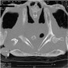
The left shoulder joint appears normal and the head of the humerus articulates with the scapula and coracoid.
Figure 2. Male radiated tortoise—transverse CT scan (bone window) at a level immediate caudal to the left shoulder
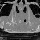
The right scapula is visible but the head of the right humerus is not.
Figure 3. Male radiated tortoise—transverse CT scan (bone window) at a level caudal to both scapulae and shoulder joints
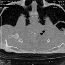
The head of the right humerus is visible, and is displaced caudodorsally from the right scapula.
Case 2
A free-living, adult, snapping turtle (Chelydra serpentina) was admitted to the Cornell University Wildlife Clinic for treatment of a fractured carapace secondary to vehicular trauma. On physical examination, the turtle was depressed, weak, anemic (PCV 10%; normal 25%), and had poor withdrawal reflexes on the left side. In the carapace, a fracture line extended approximately 7.5 cm caudally from the left cranial margin. At the cranial margin of the fracture, the shell was displaced about 6.0 mm. Radiographic evaluation revealed that the left scapula also was fractured. Computed tomography evaluation confirmed the scapular fracture and better illustrated the extent (12 mm) of displacement (Figure 4). The fracture wound was treated for 20 days and then stabilized with fiberglass and epoxy resin. The fracture was allowed to heal by second intention. Additional treatment consisted of half-strength Lactated Ringer’s solution and 2.5% dextrose (Baxter Healthcare Corporation, Deerfield, IL, USA) at 10.0 ml/kg IP, SID for 7 days, cefazolin (Ancef, SmithKline Beecham Pharmaceuticals, Philadelphia, PA, USA) at 22.0 mg/kg SC, SID for 14 days, and enrofloxacin (Baytril, Bayer Corporation, Shawnee Mission, KS, USA) at 2.5 mg/kg SC, SID for 7 days followed by 5.0 mg/kg SC, SID for the next 14 days. The anemia was treated with injections of iron dextran (The Butler Company, Columbus, OH, USA) at 12.0 mg/kg IM once weekly for 5 wk, and vitamin B complex (The Butler Company, Columbus, OH, USA) at 2.5 mg/kg of thiamin SC, SID for 7 days. The turtle was kept out of the water until the shell was repaired but daily soaking in a shallow pool was allowed. Forty-two days after the initial examination, the turtle was eating, beginning to use the left thoracic limb, and released to a wildlife rehabilitator for further care. The turtle was released to the wild 9 mo later.
Figure 4. Male snapping turtle—3-dimension CT reconstruction of the fractured left scapula