Practical Considerations for the Use of the Tubular External Fixator (F.E.S.S.A.) in the Treatment of Fractures in Birds
Abstract
This presentation describes a new model of a tubular external fixator in avian surgery. The Fixateur Externe du Service de Santé des Armées (F.E.S.S.A.) system was originally developed by the French army as a light tubular external fixator for human hand and foot fractures. Its excellent application for small bones has led to its successful use since 1990 in small animal medicine, especially in dogs, cats and rabbits with a body weight below 5 kg.2,3,5 The F.E.S.S.A. system (Figure 1) is made of stainless steel and is extremely lightweight (<10 g). Compared to other commercial external fixators the system was shown to be considerably easier to apply. Considering that the F.E.S.S.A. system can be reused in several patients, the costs favorably compare to free-form fixations such as polymethylmethacrylate, where the pins represent the major cost factor. Different models of the F.E.S.S.A. system are available with a tube diameter of 6, 8, and 12 mm. Kirschner pins of up to 2 mm and 2.5 mm respectively may be used. The length of the fixators varies from 30 mm to 118 mm. A linear or angular elongation by attachment of two fixators is possible. Minimum distance between pins is 2 mm. The pins may be placed vertically or in a 30° angle. The fixator may be used in different ways, such as a type I–III external fixator or as “tie in” fixator (combination of intramedullary pin and external fixation). At our clinics, the F.E.S.S.A. system has been used in birds with body weights ranging from 90 to 1000 g, with a success rate comparable to other avian orthopedic studies. The species where the system has been mainly used are psittacine, raptors, and pigeons to treat tibiotarsal fractures, humeral fractures, tarsometatarsal fractures, femoral fractures and antebrachial fractures. In all cases the 6-mm diameter F.E.S.S.A. system was applied with lengths varying between 30 and 70 mm and pins ranged from 0.8 mm to 1.35 mm in diameter. In mammalian surgery the use of threaded pins together with external skeletal fixation is generally recommended due to the increased pin/bone interface. In addition, positive-threaded pins are known to have a superior stability over negative-threaded pins, hence reducing the risk of pin breakage.1 In our experience the use of threaded pins can also be recommended in avian surgery; however, negative-threaded pins appear to be safe in the bird species included in the present study.
Figure 1. Schematic view of the components of the F.E.S.S.A. external fixator
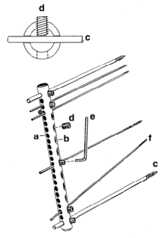
Tube with gliding (a) and threaded (b) holes, Kirschner pins (c), screws (d), and Allen key (e). Insertion of pin perpendicular (c) or at 30° angle (f).
In five out of ten ulna fixation procedures in pigeons, fissure formation or refracturing was observed. In most cases this complication occurred around the most distal pin and occurred regardless of whether the radius was additionally stabilized with an intramedullary pin or not. A possible explanation may be that the 4-cm connecting bar (including six pins) was not long enough. In birds, leverage exerted on an ulnar fracture is significantly higher than in mammals due to a different relationship of muscle insertions to joints.4 Howard (1990) recommended the use of longer plates in birds compared to similar fractures in small animals.4 This suggestion might also apply for external fixation.
In large psittacine birds it is recommended to additionally stabilize screws with a cyanoacrylate tissue adhesive (Vetbond™, 3M Switzerland), since they tend to manipulate them, which may lead to instability. In our experience the F.E.S.S.A. system is well tolerated and offers a wide range of applications in relation to the size of birds treated and the fracture types. It therefore represents a valuable alternative to currently used external fixator systems.
Literature Cited
1. Anderson, M.A., D.N. Aron, and R.H. Palmer. 1997. Improving pin selection and insertion technique for external skeletal fixation. Comp. Cont. Ed. 19: 485–494.
2. Chancrin, J.-L., T. Boubee, and M. Marguin. 1990. Utilisation du fixateur externe du service de santé des armées (FESSA) en chirurgie orthopédique vétérinaire: à propos de 29 cas. Prat. Med. Chir. Anim. 25: 217–223.
3. Haas, B., I.M. Reichler, and P.M. Montavon. 2003. Use of the tubular external fixator in the treatment of distal radial and ulnar fractures in small dogs and cats. Vet. Comp. Orthop. Traumatol. 16: 132–137.
4. Howard, P.E. 1990. The use of bone plates in the repair of avian fractures. J. Am. Anim. Hosp. Assoc. 26: 613–622.
5. Reichler, I.M., C.J. von Werthern, and P.M. Montavon. 1997. Der tubuläre fixateur externe (F.E.S.S.A.): klinische Anwendung zur Frakturversorgung bei 6 Zwerghunden und 20 Katzen. Kleintierpraxis. 42: 407–419.