Abstract
To further our understanding of hippo (Hippopotamus amphibius) biology, skin tissue samples were collected from free-ranging hippos to obtain DNA for a population genetics study. This required the development of a biopsy dart tip specifically designed for hippo skin. Samples of skin were collected from individual hippos in intact herds during the day by using a biopsy dart tip attached to a crossbow bolt. The bolt, projected from a crossbow, was retrieved via a fishing reel mounted on the crossbow with the line attached to the bolt carrying the biopsy dart. This method of collecting skin tissue samples has previously been used successfully with free-ranging whales.1,4
A whale skin tissue biopsy dart tip of simple design and successfully proven in the field on free-ranging whales1 was tested on three hippos exhibited in the Toledo Zoo’s Hippoquarium (a 360,000-gallon, filtered, underwater viewing, naturalistic exhibit) at a range of 25–30 m. Animals darted at the Toledo Zoo were individually monitored for any adverse effects to health (i.e., infection of biopsy site) by close visual inspection in the hippo restraint device in the hippo holding facility at the Toledo Zoo.2 No adverse reactions were observed. Several unsuccessful tests using the whale biopsy dart tip and modifications of this simple design were carried out. Hippo skin is composed of a thin epidermis and thick dermis layer, averaging 3.5-cm thick, 20–80 cm from the midline of the back,3 which is highly fibrous and tough. The characteristic punch, bounce, and tear mechanism of the whale biopsy dart tip simply would not work with hippo skin. A new design with movable parts that would snip and hold a piece of skin tissue upon impact was developed (Figure 1) and tested on the hippos at the Toledo Zoo. The dart tips successfully excised adequate amounts (0.25–0.5 cc) of skin tissue needed for extracting and analyzing DNA.
Figure 1. Hippo skin tissue biopsy dart: full view and cutaway; dart weight=25 grams
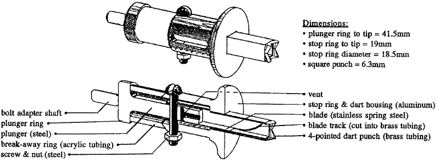
The parts for 66 dart tips were fabricated in a machine shop and were assembled in the field. All of the darts were used in the field, and some were reused after disassembly, cleaning, and reassembly. The following is a description of how the biopsy dart tip works:
1. The bolt with the attached biopsy dart tip is projected from a crossbow toward the target.
2. The four-pointed tip of the dart cuts into the skin and penetrates to the depth of the stop ring.
3. The force of the bolt pushes the steel plunger ring forward, shattering the acrylic ring (in place to prevent the plunger from pushing the cutting blade down the blade track when the bolt is projected from the crossbow) and allowing the plunger to push the stainless spring steel blade down the curved blade track.
4. The blade snips off the portion of skin inside the dart tip and closes the dart tip opening with the sample held in place.
5. The dart tip pops out of the hippo skin with the rebound of the impact.
6. The bolt with the attached biopsy dart tip and sample are retrieved with the fishing reel and attached fishing line.
The samples were then removed from the biopsy darts and placed in containers with preservatives and stored for future analysis.
The collection period was carried out during the beginning of the dry season (June–August 1997) in Kruger National Park located in South Africa. We sampled hippo herds located in narrow, shallow and accessible stretches of the Olifants and Letaba rivers at a minimum darting range of 20 m and a maximum of 45 m. When possible, hippos were darted on their sides or backs while they rested on beach areas; however, after the initial volley of beach shots, the hippos took cover in the water, usually leaving only the nape of the neck and/or back of the neck/head area for a target. This required much greater accuracy to hit the target and thus increased the percentage of missed shots. The majority of tissue samples collected were from hippos darted in the water. Care was taken to dart hippos only when they were facing away from the researcher, so as to prevent injury to the eyes, ears, or nostrils.
Our sampling of five herds (three of them with adjacent territories along the Olifants River) resulted in 56 individual samples (Table 1).
Table 1. Results of crossbow shots and hits of hippos with the skin biopsy dart
|
Hits retrieved with tissue
|
56
|
|
Hits retrieved with no tissue
|
7
|
|
Hits—line or bolt broke (lost sample)
|
11
|
|
Total hits
|
74
|
|
Missed shots
|
179
|
|
Total shots
|
253
|
Percentage of total shots that were hits: 29%
Percentage of total shots retrieved with tissue: 22%
Percentage of retrieved hits with tissue: 89%
The basic design and the results of field testing of the hippo skin biopsy dart were successful and may be applicable for use with other thick-skinned mammals such as the rhinoceros (W. Karesh, The Wildlife Conservation Society, New York, NY, personal communication).
Additional Notes
The crossbows used were both made by Hunter’s Manufacturing Company, Inc. The Huntmaster Advantage model with 68-kg draw weight was used for the tests at the Toledo Zoo, and the 458 Magnum Treestand with a 75-kg draw weight was primarily used in the field.
The spool on the open-face spin cast fishing reel (mounted on the top front end of the crossbow just behind the bow limbs) was filled with either 23- or 27-kg test SpiderWire, high tensile strength microfilament braided fishing line, tied to a double loop of 36-kg SpiderWire attached to the butt end of the bolt. The leader of the line was laid down in the groove below the bolt track from front to back. All knots in the SpiderWire were secured with superglue.
A safety line, in the event of bolt breakage upon impact, was attached from the loop on the butt end of the bolt to the dart tip.
Acknowledgments
Disney’s Animal Kingdom for primary funding of this project. Apex Design and Manufacturing Inc. for funding, designing, and fabricating of the biopsy dart and the crossbow fishing reel mounts. Kruger National Park for providing lodging and field logistics. The Toledo Zoo for allowing us to test the biopsy darts on the hippos exhibited in the Hippoquarium. Alex Krajcirovic for mechanical engineering of the biopsy dart. Moira Brown for advice concerning the design of biopsy darts and delivery systems. William Karesh, Timothy Reichard, and Wynona Shellabarger for advice on the design of biopsy darts. The employees of Cleland’s Outdoor World for expert advice on all matters about crossbows and archery. Finally, we thank our partners, Mary Beth McConnell, Lori-Ann LeBlanc, and Patricia Stilwill for their support and invaluable assistance with this project.
Literature Cited
1. Brown, M.W., S.D. Kraus and D.E. Gaskin. 1991. Reaction of North Atlantic right whales (Eubalaena glacialis) to skin biopsy sampling for genetic and pollutant analysis. Reprint of the International Whaling Commission. Special issue 13: 81–89.
2. Krueger, S., W. Shellabarger and T. Reichard. 1996. Hippopotamus training: implications for veterinary care. In: Proceedings of the 1996 American Association of Zoo Veterinarians Annual Conference. pp. 54–58.
3. Luck, C.P. and P.G. Wright. 1964. Aspects of the anatomy and physiology of the skin of the hippopotamus (H. amphibius). Quarterly Journal of Experimental Physiology. 49(1): 1–14.
4. Palsbøll P.E., F. Larsen and E.S. Hansen. 1991. Sampling of skin biopsies from free-ranging large cetaceans in West Greenland: development of new biopsy tips and bolt designs. Reprint of the International Whaling Commission. Special issue 13: 71–79.