Epulides/epuli (plural of epulis) are common benign growths found in dogs' mouths.
There are three types:
Epulis, Dog (Photograph)
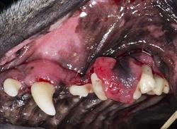
Fibromatous Epulis was confirmed with biopsy. Courtesy Dr. Jan Bellows
Fibromatous epulis appears on a stalk of tissue, much like a mushroom, or as an unmoving mass. It is usually pink in color and has a non-ulcerated smooth surface. It may appear as an enlargement on the gum tissue near incisor, canine, or premolar teeth.
Peripheral odontogenic fibroma (previously called ossifying epulides) is similar in appearance to a fibromatous epulis as it also has a pink smooth surface, but it has an osteoid matrix; it's made up of early-stage bone cells known as osteoblasts.
Acanthomatous ameloblastoma (previously called acanthomatous epulis) is classified as benign but it tends to invade adjacent bone and it is locally aggressive. It can be pre-cancerous. However, it does not spread to other regions of the body. This tumor often has a rough cauliflower-like, ulcerated surface. It occurs most commonly in the incisor and canine tooth area of the lower and upper jaw. It occurs less commonly near the fourth upper premolar in the upper jaw and the lower jaw's first molar.
Statistics
Epulis is the fourth most common tumor found in the canine mouth. It is very rare in cats.
This tumor is more common in brachycephalic breeds. Brachycephalic breeds are those that have a very short nose and muzzle. Boxers and English Bulldogs are brachycephalic dog breeds, for example.
Middle-aged and older dogs get epulides more often than young dogs do. The average age of an affected dog is seven years old.
The overgrowths are generally a reaction to trauma, such as a tooth rubbing on the gum in brachycephalic mouths, for example.
Clinical Signs
Signs include a lump on the gums, drooling, halitosis, facial deformity, and other signs of mouth injury. The visible lump is the most common sign.
Diagnosis
Diagnosis begins with a visual examination of the mouth.
Radiographs of the head can determine how invasive the tumor is.
Biopsy and histopathological (microscopic) examination of the lump will determine which type of epulis is present.
Treatment
Fibromatous epulis: Treatment involves removing the mass, extracting the involved tooth, and thoroughly scraping the tooth socket clean.
Peripheral odontogenic fibroma: Treatment involves removing the mass, extracting the involved tooth, and thoroughly scraping the tooth socket clean. This surgery can be more difficult than that for fibromatous epulis.
Acanthomatous ameloblastoma: Treatment is surgical removal, including removing the affected areas of the upper or lower jaw (maxillectomy or mandibulectomy).
In some inoperable epulis cases, radiation therapy may help.
Aftercare
After surgery, your pet may need a softer diet. Your veterinarian will advise you about this.
Prognosis
The less tissue involved, the better. If a large section of the jaw has to be removed, it can affect the dog's quality of life.
The oral tumors don't usually recur if the entire tumor has been removed. However, some can. Checking your dog's mouth frequently will help you spot any tumor recurrence or new tumors.