Thromboelastography Guided Diagnosis and Therapy in a Case of Elephant Endotheliotropic Herpesvirus Hemorrhagic Disease
Abstract
Elephant endotheliotropic herpesviruses can lead to a devastating hemorrhagic syndrome (EEHV HD) in sub-adult elephants with an extremely high mortality rate. Clinical symptoms result from reduced endothelial integrity leading to widespread edema, hemorrhage and presumptive consumption of platelets and clotting factors. These hemostatic abnormalities have not yet been characterised. Thromboelastography (TEG) measures the dynamic process of coagulation and fibrinolysis1 and has numerous applications in veterinary species2.
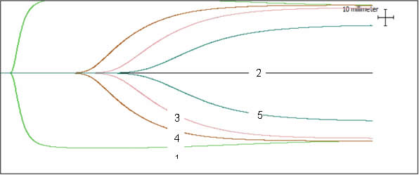
A 20-mo-old Asian elephant (Elephas maximus) presented with non-specific signs. TEG performed on citrated whole blood revealed a hypocoagulable state (Figure 1, line 2) which supported suspicions regarding EEHV HD. Aggressive treatment included whole blood and plasma transfusions, antiviral medications (ganciclovir and famciclovir), synthetic factor VII (NovoSeven, NovoNordisk Scandinavia AB, Region Denmark, DK-2300, approximately 70 μg/kg IV) and supportive care. Repeat TEG showed some improvement (Figure 1, line 3). Initially the R value, indicating time to initial fibrin formation, was greater than 119 minutes, but decreased to 43.3 minutes after treatment. R in normal elephants is 3–4 minutes. Despite treatment, the calf deteriorated and was euthanized. Pathology and PCR confirmed a diagnosis of EEHV-1A.
In vitro analyses were performed to assess the potential effect of 20 and 40 ml/kg plasma therapy (Figure 1, lines 3 and 4, respectively). However in vivo, this would require administration of 15–30 L of plasma. As less than 1200 ml of plasma were successfully given, it is believed that the improvement seen on TEG was associated with administration of synthetic factor VII.
Figure 1

TEG curve from 17-year-old healthy dam (1); EEHV HD calf pre-treatment (2 = straight line); in vitro addition of 50 µl (3) and 100 µl (4) plasma from dam to calf sample; calf sample after synthetic factor VII treatment (5).
TEG shows potential to characterise and assess the pathogenesis and potential treatment options in elephants with EEHV HD.
Acknowledgments
The authors would like to thank the keeping staff at the Copenhagen Zoo, Louise Bochsen and Gitte Hansen for their support with this case.
Literature Cited
1. Whiting D, DiNardo JA. TEG and ROTEM: technology and clinical applications. Am J Hematol. 2014;89:228–232.
2. Winberg B, Kristensen AT. Thromboelastography in Veterinary Medicine. Semin Thromb Hemost. 2010;36:747–756.