Dennis J. Chew1, DVM, DACVIM (Internal Medicine); Patricia A. Schenck2, DVM, PhD
An increased serum calcium is typically first noted when total calcium (tCa) is measured as part of a biochemistry profile. Abnormalities in tCa warrant further diagnostic investigation. First it should be verified that the abnormality is repeatable. If the abnormality is repeatable, ionized calcium (iCa) should be measured for an accurate assessment of calcium status. Total or adjusted tCa are not reliable measurements of calcium status as noted by a high degree of diagnostic discordance between total, adjusted, and ionized calcium measurements.
Small increases in ionized serum calcium concentration can have adverse consequences in some animals whereas others with a similar or greater degree of hypercalcemia may not manifest obvious clinical signs. A mild degree of hypercalcemia may not be immediately dangerous and there is time to establish a definitive diagnosis before starting treatment. In those with severe clinical signs associated with hypercalcemia, diagnostic and therapeutic efforts may need to proceed concurrently. Interaction with serum phosphorus is important, as those with a tCa (mg/dL) times phosphorus concentration product greater than 70 are most likely to have severe tissue changes associated with mineralization. Hypercalcemia can be toxic to all body tissues, but major deleterious effects are on the kidneys, nervous system, and cardiovascular system. Most animals with tCa greater than 15.0 mg/dL will show systemic signs, and those with tCa concentrations greater than 18.0 mg/dL are critically ill.
Polydipsia, polyuria, and anorexia are the most common clinical signs attributed to hypercalcemia, though depression, weakness, vomiting, and constipation can also occur. Uncommonly, cardiac arrhythmias, seizures, and muscle twitching are observed. Severe hypercalcemia that has developed rapidly (hypervitaminosis D) can result in death. Cats with hypercalcemia do not display polyuria, polydipsia or vomiting as commonly as do dogs with a similar degree of hypercalcemia. Cats with idiopathic hypercalcemia may have no obvious clinical signs.
Differential Diagnoses for Hypercalcemia
Hypercalcemias can be classified as parathyroid-dependent (primary hyperparathyroidism), or parathyroid-independent (normal parathyroid gland). In hypercalcemic dogs, neoplasia is the most common diagnosis, followed by hypoadrenocorticism, primary hyperparathyroidism, and chronic renal failure. Approximately 70% of hypercalcemic dogs are also azotemic, with azotemia uncommon only in dogs with hyperparathyroidism. In hypercalcemic cats, neoplasia is second to renal failure or idiopathic hypercalcemia.
 H = Hyperparathyroidism (1°, 3°, hyperplasia), humoral hypercalcemia of malignancy, houseplants, hyperthyroid
A = Addison's disease, aluminum toxicity, vitamin A, milk-alkali
R = Renal disease, raisins (grapes)--dogs
D = Vitamin D toxicosis (granulomatous Dz), drugs, dovonex, dehydration, DMSO (calcinosis cutis), diet
I = Idiopathic (cats), infectious, inflammatory, immobilization
O = Osteolytic (osteomyelitis, immobilization, local osteolytic hypercalcemia, bone infarct)
N = Neoplasia (HHM and LOH), nutritional
S = Spurious, schistosomiasis , salts of calcium, supplements
Idiopathic Hypercalcemia of Cats
A syndrome in young to middle-aged cats has emerged, where hypercalcemia occurs without obvious explanation. Serum total calcium is increased for months to more than one year, often without obvious clinical signs. Ionized calcium is increased, sometimes out of proportion to the increase in total serum calcium. Longhaired cats may be over represented in this syndrome. Vomiting and weight loss are the most common clinical signs. Most are nonazotemic, but may develop azotemia at a later date. Nephrocalcinosis is occasionally observed, as are uroliths in the kidney, ureter, and bladder. There is no evidence of malignancy based on radiography, abdominal ultrasonography, bone marrow evaluation, and in some instances, full necropsy. Serology for FeLV and FIV is negative, and T4 values are normal. PTH levels are within the reference range, PTHrP is not detectable, and 25-(OH)-D and calcitriol levels are within normal limits. Blood gas analysis reveals no major acid-base disturbance.
An increase in dietary fiber has been reported to decrease serum calcium in affected cats in some reports. Challenge with prednisone therapy results in long-term decreases in iCa and tCa in some cats. There is concern that this treatment could increase hypercalciuria, which could subsequently enhance genesis of urinary calculi. However, the declining filtered load of calcium decreases as serum iCa declines, which offsets the enhanced formation of calculi. When dietary modification and challenge treatment with prednisolone have been unsuccessful in resolving hypercalcemia, bisphosphonate treatment should be considered. The cause(s) of idiopathic hypercalcemia in cats remains elusive. The role of dietary acidification, dietary magnesium restriction, and/or contribution of any specific dietary constituents deserve further consideration. It is conceivable that hypercalcemia develops only in a genetically susceptible population of cats.
Click on the image to see a larger view
| | 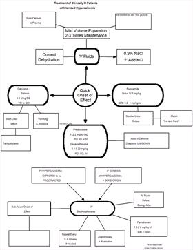
Treatment algorithm for consideration in patients that have ionized hypercalcemia and are obviously sick. |
|
| |
|