Dental disease is prevalent among domestic cats. Approximately 70% of cats over the age of three years suffer from some degree of dental disease. However, clients may not be aware of a problem until the disease has reached an advanced stage. Oral inspection during annual physical examination may reveal evidence of periodontal disease, feline odontoclastic resorptive lesions, or endodontic disease. Dental radiography is essential in order to more accurately diagnose these conditions, to formulate a therapeutic plan, and to assess therapeutic success (i.e., during extractions or endodontic obturation).
While it is possible to obtain dental radiographs with extra-oral film and standard radiographic equipment, the use of intra-oral radiographic film and a dental x-ray unit provides superior images with minimal radiation exposure. However, conventional radiographic techniques often result in superimposition of anatomical structures and may provide unsatisfactory views of individual teeth. In addition, cats present a challenge because of their compact skull shape, and it is often difficult to obtain satisfactory views of the maxillary cheek teeth due to the overlying zygomatic arch.
Techniques for obtaining diagnostic dental radiographs have been described in several veterinary journal articles and dental textbooks. Despite this, obtaining consistently good radiographs of feline maxillary premolar teeth has proved to be difficult. The extra-oral, near-parallel technique provides a repeatable method for achieving high-quality radiographs of the maxillary premolars and molars.
STANDARD RADIOGRAPHIC VIEWS IN THE CAT
Six views are adequate for visualization of the entire feline dentition. When taken by an inexperienced student assisted by an experienced veterinary dental technician or clinician at the University of California Veterinary Medical Teaching Hospital, a set of radiographs is completed in approximately thirty minutes. With practice, full-mouth radiographs of consistent quality may be obtained in fifteen minutes or less. The following table summarizes the recommended radiographic views in the cat:
|
View |
Position |
Technique |
Film Size |
Exposure Time
(60 kV, 7 mA) |
|
maxillary I and C |
intra-oral (occlusal) |
bisecting angle (C) |
# 4 |
0.16 s |
|
maxilla: P2—M1 |
extra-oral |
near-parallel |
# 2 |
0.20 s |
|
mandibular I and C |
intra-oral (occlusal) |
bisecting angle (C) |
# 2 |
0.12 s |
|
mandible: P3–M1 |
intra-oral |
parallel |
# 0 or 2 |
0.12 s |

|
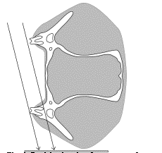
Fig1: Positioning for the extra-oral near-parallel view of the maxillary fourth premolar in the cat |
|
| |
Maxillary premolars and molar: Image overlap of the zygomatic arch and maxillary premolars commonly occurs in the cat. In order to avoid this problem, the extra-oral near-parallel technique is recommended (Fig. 1). Any films used extra-orally must be marked to separate them from intra-orally exposed film to ensure proper orientation when processed.
LATERAL VIEW OF THE CANINES
The occlusal views of the maxillary and mandibular canines and incisors provide satisfactory visualization of these teeth for routine evaluation. In cases where pathology of the canine teeth is suspected, however, these views are inadequate due to superimposition of the canines and the premolars, obscuring the apices of the canines. The lateral view is recommended when evaluation of the periapical region of the canine tooth is required. A bisecting angle technique is employed, using the long axis of the canine tooth as viewed from the front of the cat (Fig. 2). The same technique may be used for the mandibular canines.
ROUTINE USE OF FULL-MOUTH RADIOGRAPHS IN CATS
Dental radiography forms an integral part of feline practice. Most common dental conditions, including periodontal disease and endodontic disease, develop in cats and are associated with radiographic signs. The value and routine use of a full-mouth radiographic survey taken during a (human) patient’s first visit to a dentist is well established. Routine use of full-mouth radiography would appear to be more common in cats than dogs. One feline practitioner reported that full-mouth survey radiography accounted for 42% of all radiographs taken in the practice. The increasing sophistication of veterinary dentistry and increased availability of suitable dental radiographic equipment, raises the question of whether the current standard of care should be upgraded to include full-mouth radiography when the patient is first examined for dental treatment.
The objective of a recent study at UC Davis was to evaluate the relation between presenting dental complaint, clinically observed oral disease indicators, and radiographic findings on full-mouth radiographs. The study was undertaken in a series of 115 cats referred for dental treatment and for which full-mouth radiography had not been done previously.
The main clinical findings were radiographically confirmed in all cats. Periodontal disease was the presenting complaint and a main clinical finding in the majority of cats. Radiography provided additional information on the extent of the expected supporting bone loss in 92.7% of cats with periodontal disease. In 25.2% of all cats, radiography revealed teeth with the extent of periodontal disease and bone loss greater than what was clinically diagnosed or expected. The odds ratios between periodontal disease and chronic stomatitis as presenting complaint or main clinical finding and the presence of root fragments, were significantly increased.
External odontoclastic resorption lesions were diagnosed clinically in 61 (53%) cats. In 98.4% of these cats, radiography revealed additional information on extent of the lesions. Only in 8.7% of cats were external odontoclastic resorption lesions seen on radiographs but were not clinically evident. Evidence of root resorption and ankylosis was seen in 41 cats (35.7%), and the odds ratio for this finding was significantly increased if a diagnosis of odontoclastic resorption lesions was made. Internal resorption, without signs of external resorption, was seen in only one cat.
In all, 87 cats (75.5%) had missing teeth as a main clinical finding. The odds ratio of finding root fragments was significantly increased in cases of chronic stomatitis and severe periodontal disease. Additional radiographic information indicated 25.3% of cats had no evidence of root tips or alveolus. However, 72.4% of cats with missing teeth had evidence of root tip fragments and 2.3% of cats had radiographic evidence of embedded teeth.
Endodontic disease (complicated tooth fractures, crown discolouration) was a main clinical finding in only 14 cats. Periapical lesions were present in 42.9% of these cats.
The unexpected radiographic findings include developmental conditions: supernumerary roots and conditions, such as periapical lesions (without clinical evidence of endodontic disease) and root resorption, for which clinical signs of disease are not manifested:
|
Prevalence of unexpected radiographic findings |
|
Finding |
Frequency |
|
Root resorption/ankylosis
Extent of periodontitis/bone loss
Odontoclastic resorption lesion
Root fracture
Periapical lesion
Supernumerary tooth
Supernumerary root
Periodontal-endodontic lesion |
35.6%
25.2%
8.7%
7.0%
3.5%
1.7%
1.7%
1.7% |
The value of the radiographs in clinical decision-making was analysed and the results depicted in the above piecharts. The assessment of the clinical value of radiographs of teeth with clinical lesions is seen on the left, while the assessment of the clinical value of radiographs of teeth without clinical lesions is seen on the right.
The value of full-mouth radiographic surveys was also assessed according to the breed and age groups. Differences between the breed groups were minor and not significant for radiographs of teeth with and without lesions, respectively, yet the difference between age groups was marked. The percentage of cats in which the additional radiographic information obtained from teeth with clinical lesions was considered essential, increased from 18.2% to 21.4 and 53.5% in the three age groups (p = 0.0012). Similarly, the number of cats with clinically important unexpected radiographic findings increased from 18.8% to 35.5 and 58.8% with increasing age (p = 0.044).
SUMMARY
In summary, statistical analysis of the survey results revealed certain clinically predictable and useful patterns for cats with severe periodontal disease, odontoclastic resorption lesions, or chronic stomatitis. The odds of finding an unexpected amount of bone loss, root resorption and ankylosis, and root fragments were significantly increased. The underestimated extent of the periodontal disease is of clinical importance, especially taking into account that these patients were subjected to meticulous periodontal charting. Assessment of the clinical value of radiography in this study suggests that the diagnostic yield of full-mouth radiography in cats is high. Older cats derive markedly more benefit of full-mouth radiography than do younger cats. Larger case series would be required to investigate the effect of breed on the value of full-mouth radiography.
REFERENCES
1. Verstraete F.J.M., Kass P.H., Terpak C.H. 1998 Diagnostic value of full-mouth radiography in cats. American Journal of Veterinary Research 59: 692-695
2. Lommer M.J., Verstraete F.J.M., Terpak C.H. 2000 Dental radiographic technique in cats. Compendium on Continuing Education for the Practicing Veterinarian 22(2): 107-117
3. Lommer M.J., Verstraete F.J.M. 2000 Prevalence of odontoclastic resorption lesions and periapical radiographic lesions in cats: 265 cases (1995-1998). Journal of the American Veterinary Medical Association 217: 1866-1869
4. Lommer M.J., Verstraete F.J.M. 2001 Radiographic patterns of periodontitis in cats: 147 cases (1998-1999). Journal of the American Veterinary Medical Association 218: 230-234
5. Figures 1 and 2 are modified and reprinted with permission from: Lommer M.J., Verstraete F.J.M., Terpak C.H. 2000 Dental radiographic technique in cats. Compendium on Continuing Education for the Practicing Veterinarian 22(2): 107-117