Pathology and Epidemiology of Neoplasia in the Captive Population of Black-Footed Ferrets (Mustela nigripes)
Abstract
The capture of the last 18 known wild black-footed ferrets (Mustela nigripes) in 1986–1987 marked the beginning of one of the most successful captive propagation efforts in the history of endangered species conservation. Since then, close to 2000 black-footed ferrets have been born in the seven institutions involved in this breeding program. Neoplasia is an important cause of morbidity and mortality in ferrets after they reach adult age. In order to better characterize these neoplastic conditions, and to evaluate their significance in the management of this species, we conducted a retrospective study of the cases of neoplasia diagnosed in the captive population between 1988 and 1998. Prior to April 1, 1998, a total of 206 adult black-footed ferrets (at least 1-yr-old) have died in captivity. At this time, postmortem reports and embedded tissues were recovered from 180 of these adult ferrets (102 males and 78 females). Pertinent information was also recovered from the studbook maintained at the USFWS National Black-Footed Ferret Conservation Center. Each tumor was examined by the same pathologist, and classified according to its histologic features. A total of 172 neoplasms was diagnosed in 97 of the 180 adult ferrets (53.9%) included in this study. Multiple neoplasias were common; thirty-five, ten, four, and two ferrets being affected by respectively two, three, four, and five different types of tumors each. The prevalence of most of the tumors increased with the age at death (Figure 1). With the exception of two cases, all ferrets with tumors were older than 4 yr. Neoplasia was believed to have contributed to the death of 59 ferrets (32.8%), all but two being over 4-yr-old (Figure 1). The most common tumors observed were, in decreasing order of frequency, renal tubular neoplasms (prevalence: 21.7%); sweat gland adenomas/adenocarcinomas (prevalence: 19.4%); biliary cystadenomas/cystadenocarcinomas (prevalence: 14.4%); mammary adenocarcinomas (prevalence in females: 15.4%); adenomas/adenocarcinomas of the perianal glands (prevalence: 8.3%); adenomas/adenocarcinomas of the preputial glands (prevalence in males: 5.9%), and squamous cell carcinomas (prevalence: 4.4%). At least 14 other types of tumors were also present.
Figure 1. Age distribution of biliary cysts and various neoplasms in postmortem examination of adult (>1-yr-old) black footed ferrets, 1988–1998
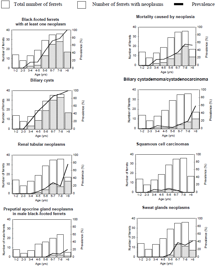
n=171 (age was available for 98 males and 73 females)
Renal tubular neoplasms were commonly multiple and bilateral. These usually protruded from the renal capsule and ranged from small, microscopic aggregates of neoplastic tubular cells to large locally invasive masses replacing most of the renal parenchyma. Based on their cellular morphology and invasive behavior, most of these tumors were classified as renal carcinomas, but distant metastases were found in only one of the 39 cases.
Biliary cystadenocarcinomas were usually fast growing, markedly invasive malignancies, frequently associated with extensive abdominal carcinomatosis. An interesting morphologic feature of these tumors was their usual affiliation with large biliary cysts. Neoplastic papilliform growths could usually be seen originating from the epithelium lining these cysts. Biliary cysts were very common in the liver of adult black-footed ferrets (65.6% of the animals examined). These cystic dilations of bile ducts seem to increase in size and in number with age. Their etiology is at this time unclear.
Most of the sweat gland tumors were cystic, slow growing, and located on the tail. Despite the anaplastic appearance of the cells in most of these tumors, distant metastases were rarely observed. In contrast, adenocarcinomas of the perianal apocrine glands were usually markedly invasive, and frequently metastasized to regional lymph nodes, abdominal cavity and lungs. These tumors were mainly observed in males. Three of the apocrine preputial gland tumors were classified as adenomas and three as adenocarcinomas. Abdominal metastases were present in one of these cases. All the 12 tumors of the mammary glands diagnosed were classified as adenocarcinomas, and distant metastases were detected in four of these cases.
Most squamous cell carcinomas affected the oral cavity, where they were markedly invasive. Pulmonary metastases were detected in one animal.
This study shows that neoplasms, especially of epithelial origin, are very common, and represent an important cause of mortality in captive adult black-footed ferrets. The very high prevalence of neoplasia in this population is intriguing, and suggests a potential genetic predisposition, and/or exposure to carcinogenic factors.
The impact of a high prevalence of neoplasia on the captive propagation of this species is most likely limited, since tumors are encountered almost exclusively in post-reproductive ferrets. The effect on the wild population would also probably be insignificant, since ferrets released into their natural habitat rarely reach the age when these neoplasms occur.
Acknowledgments
Funding for this project, and the stipend for S.L., was generously provided in full by the Zoological Society of Toronto. We are grateful to clinicians and pathologists involved in the black-footed ferret captive propagation program, including Corrine Brown (Omaha’s Henry Doorly Zoo), Roy Burns (Louisville Zoo), Graham Crawshaw (Toronto Zoo), Mary Duncan (St. Louis Zoological Park), Della Garell (Cheyenne Mountain Zoo), Julie Kreeger (National Black-Footed Ferret Conservation Center), Don Kwiatkowski (formally at the National Black-Footed Ferret Conservation Center), Dick Montali (National Zoological Park), Jon Patterson (Michigan State University), Allan Pessier (National Zoological Park), and Chris Schiller (All Creatures Pathology Service). Our thanks also goes to Bill Russell for studbook keeping, and to Paul Marinari and Astrid Vargas from the USFWS National Black-Footed Ferret Conservation Center.