Abstract
MRI employs powerful magnetic fields and radio frequency electromagnetic energy to produce images. Like computed tomography (CT) scans the output of MRI scans is digital and can be extensively post-processed. Unlike CT, these image data largely reflect the behavior and position of protons within water molecules in the tissues. Images produced by MRI scans of postmortem specimens are extremely valuable as reference materials to guide the use of other imaging modalities. A poster presentation does not allow for dynamic post-processing of these data such as three-dimensional reconstruction, digital subtraction techniques, etc. These techniques are perhaps the most powerful application of MRI data sets and should be pursued in marine mammal species.
Introduction
Magnetic resonance imaging (MRI) is the medical application of nuclear magnetic resonance spectroscopy or NMR. The technique has its origin in structural determination methods of organic chemistry. MRI employs powerful magnetic fields and radio frequency electromagnetic energy to produce images. All nuclei with odd-numbered masses or even-number masses but odd atomic numbers exhibit magnetic properties. That is they behave as though spinning about an axis. Normally the orientation of the spin, and therefore, magnetic moment, is random. However, when subject to strong enough magnetic fields the spins will align. If while aligned the nuclei are exposed to appropriate radio frequency electromagnetic pulses they will absorb energy and achieve “resonance” with the frequency. These resonance shifts can be detected by sensitive radio frequency receivers and mapped to the image space as it corresponds to their position. Like computed tomography (CT) scans the output of MRI scans is digital and can be extensively post-processed. Unlike CT, the image data largely reflect the behavior and position of protons within water molecules in the tissues.
The use of MRI in marine mammal investigations has been limited largely to experimental use with postmortem specimens.1-3 The constraints of magnet size and location, patient size, restraint, and the unique requirements of marine mammals away from an aquatic environment seriously limit the clinical use of this technology. However, images produced by MRI scans of postmortem specimens are extremely valuable as reference materials to guide the use of other imaging modalities.
Methods
A whole-body MRI data set was collected 5 hours postmortem from an 8-year-old, 237 cm, 150 g female Atlantic bottlenose dolphin, (Tursiops truncatus). Images were produced using a General Electric Medical Systems, Genesis Signa diagnostic unit with T2 weighted technique. The animal was positioned on the magnet gantry in left lateral recumbency. Two perpendicular scan planes were used and their orientation to the animal’s axis is shown graphically in Fig. 1.
Figure 1
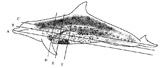
Scan planes for MRI data set collected from Atlantic bottlenose dolphin.
Three dorsal plane images and three transverse plane images demonstrating clinically relevant anatomy have been selected, digitally reproduced and are displayed. The scan planes for these images are shown in Fig. 1. On the poster dorsal plane scans are oriented inferior to superior, left to right respectively and transverse plane images are oriented rostral to caudal, upper to lower respectively. Clinically relevant anatomy is identified by alphabetic key posted to the left of the images.
Results
Dorsal Plane Images
Image A visualizes the intrathoracic anatomy at the level of the heart base and also includes the cranial sector of the abdomen. The spatial relationship of the pyloric chamber, pancreas, spleen, forestomach, fundic chamber, and small intestine is clearly imaged and of particular clinical value.
Image B clearly shows the intrathoracic anatomy at the level of the primary bronchi and the cranial sector of the abdomen at the level of the fundic chamber and forestomach. Dependant fluid accumulation is seen in the left aspect as increased signal strength. The animal was positioned in left lateral recumbency during the immediate postmortem period and scan acquisition. Recently ingested fish is visible within the forestomach lumen.
Image C demonstrates the cranial and middle sectors of the abdomen. Note the ventral flexure of the pyloric chamber, the pancreas, spleen, and the position of both kidneys.
Transverse Plane Images
Image D shows the intrathoracic anatomy at the level of the heart. Dependant fluid accumulation is again evident as increased signal strength. Also note the compression of the dependent left lung and the position of the margin of both lungs with respect to the heart. Recently ingested fish is seen within the esophageal lumen.
Image E demonstrates the overlap of the caudal sector of the thorax with the cranial sector of the abdomen. This is of particular clinical importance when utilizing other imaging techniques. Reverberation artifact during sonography reduces the acoustic window to the cranial sector of the abdomen and summation sign in radiographs will often obscure caudal pulmonary anatomy.
Image F illustrates the complex gastrointestinal anatomy of the cranial sector of the abdomen. The ampulla of the duodenum is visible dorsal to the ventral flexure of the pyloric chamber and distinguished from the later by the presence of lumenal folds. This is also clearly evident on sonography. A dependant fluid line is seen within the ampulla. Lumenal folds are also clearly visible within the esophagus as it enters the forestomach.
Discussion
Presentation of MRI images as a poster is valuable as a reference tool to guide the clinical performance and interpretation of images produced with other clinical diagnostic techniques such as radiology and ultrasonography. A poster presentation does not allow for dynamic post-processing of these data such as three-dimensional reconstruction, digital subtraction techniques, etc. These techniques are perhaps the most powerful application of MRI data sets and should be pursued in marine mammal species.
Literature Cited
1. Endo H, et al. MRI examination of trachea and bronchi in the Ganges river dolphin (Platanista gangetica). J Vet Med Sci. 1999;61(10):1137–1141.
2. Hillmann DJ. Anatomy of the fetal bowhead whale (Balaena mysticetus) using magnetic resonance images. In: Proceedings of the International Association for Aquatic Animal Medicine. 1991:172.
3. Matassa K, Early G, Wyman B, Rommel S, Krum H. The stranding of a neonate male pilot whale (Globicephala melaena); a case study. In: Proceedings of the International Association for Aquatic Animal Medicine. 1994:166.