Abstract
Bonobos (Pan paniscus) are highly intelligent, endangered apes whose natural home range is limited to the lowland rain forest south of the Congo River. In 2001, the International Studbook for bonobos listed the world population of captive bonobos as 141 animals held at 17 institutions, with 16 animals residing at the Milwaukee County Zoo. Bonobos have a low fecundity rate, with singleton births occurring at an average interbirth interval of 4 years.3 Between January 1990 and March 2000, over 30% of captive bonobo mortalities reported to the International Studbook occurred in perinatal animals. Of these deaths, 69% were due to abortion, stillbirth or congenital defect. In bonobos over 1 year of age, 46% of mortalities were associated with cardiovascular disease. Cardiovascular disease decreases the reproductive success of the captive population by affecting middle-aged animals, often of prime breeding age, with deaths reported in animals 11–29 years of age (Clyde, 2000 Veterinary Advisor Report to the Bonobo Species Survival Plan).
Ultrasonography is a valuable tool for detecting fetal abnormalities, monitoring gestation, screening animals for cardiovascular disease, and assessing clinical progression and treatment effectiveness in affected animals. The operant conditioning program for bonobos initiated at the Milwaukee County Zoo in 1993 has allowed repeated cardiac and reproductive ultrasonographic evaluations of awake bonobos in order to document baseline data for individual animals, and to develop reference parameters for the species. Measurements from a single gestation have been published previously.4
Ultrasound examinations are performed with bonobos stationed in an overhead transfer chute made of 5x5-cm metal mesh. The chute connects two play areas, and the animals frequently relax in this chute. To facilitate examinations, animals are positioned overhead in sternal recumbency with arms extended over their head for operator safety. For cardiac evaluations, animals with barrel-shaped chests are trained to twist onto their left hip in order to lower the left chest wall closer to the mesh. A 10x10-cm sliding door was designed to allow better access with the ultrasound probe, but most measurements are taken with the probe inserted through the mesh.
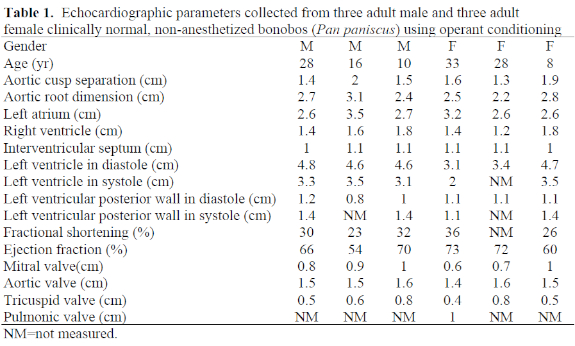
Ultrasound examinations are performed by registered ultrasonographers, and later evaluated by a consulting board-certified medical cardiologist, radiologist, or obstetrician. Echocardiograms have been collected using either a Phillips SD 800 or Phillips 2000 ultrasound machine and a phased array 2.5-MHz probe. A complete cardiac study utilizes two-dimensional (2D) images to assess the anatomic relationship and motion of the heart structures, M-mode unidimensional images for cardiac measurements, Doppler analysis to assess blood flow through the heart, and echophonocardiography for the evaluation of heart sounds and murmurs. Cardiac parameters for three adult male and three adult female clinically healthy bonobos are presented in Table 1.
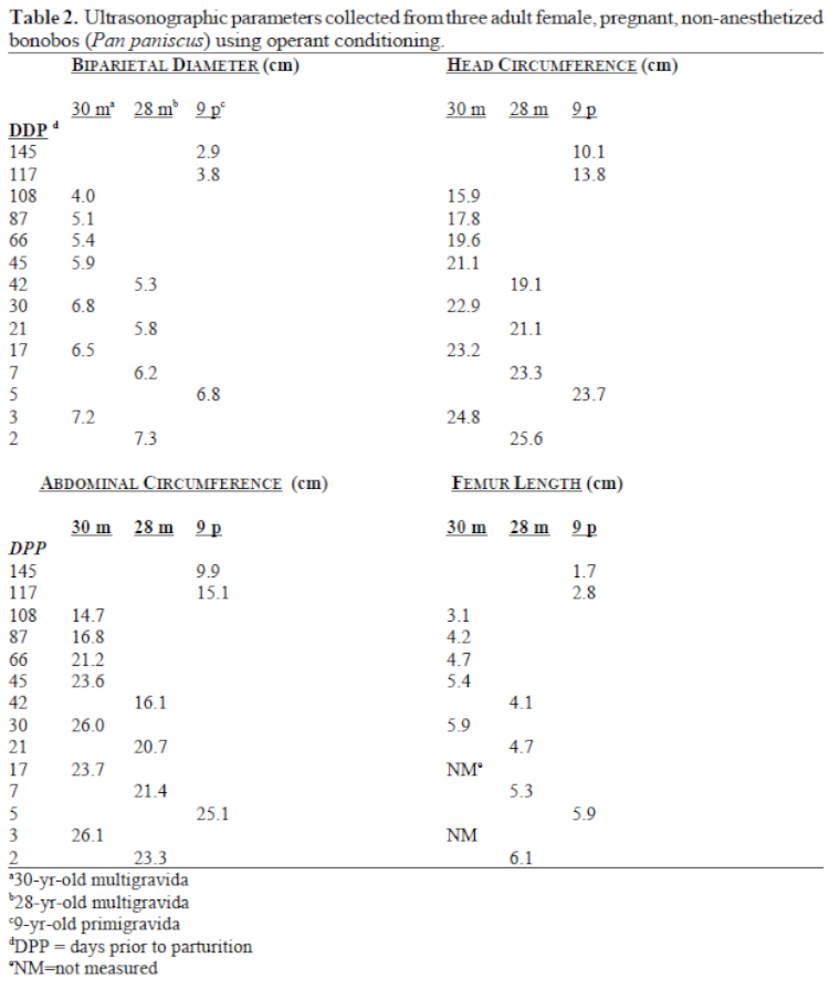
Gestational ultrasounds are performed with the same machines and probe. A complete gestational study includes detection and measurement of the fetal heart rate to verify viability, measurement, and identification of fetal structures to evaluate fetal growth and to check for possible fetal defects, assessment of fetal presentation and position, description of placental location and quality, and evaluation of amniotic fluid volume. Ultrasound parameters collected during a single nonpathologic gestation for each of three different female bonobos are presented in Table 2.
Transthoracic echocardiography and transabdominal gestational ultrasonography are effective tools for noninvasive assessment of cardiac and fetal health in the bonobo. The ability to obtain these measurements without anesthesia should allow for more frequent evaluations in this endangered species. The anatomic structure and function of the bonobo heart, placenta and fetus are very similar to humans, affording ease of interpretation for consulting medical specialists. Echocardiographic parameters measured in mature bonobos appear similar to values reported for adult humans,1 even though bonobos weigh considerably less. However, fetal parameters and fetal growth rate in bonobos are different from published human reference ranges.2 Established calculations for human fetal age appear to overestimate the age of the second-trimester bonobo fetus, while underestimating bonobo fetal age in the third trimester. Continued effort is needed from all institutions holding bonobos to collect cardiac and gestational ultrasound measurements to develop improved reference ranges in this highly endangered ape.
Acknowledgments
The success of the bonobo operant conditioning program at the Milwaukee County Zoo is due to the time, effort and assistance of the entire keeper staff of the Sterns Family Apes of Africa and Primates of the World, Curator of Primates Jan Rafert, veterinary technicians Margaret Michaels and Joan Maurer, training consultants Shelley Ballman of Oceans of Fun, Inc. and Beth Trczinko, and previous veterinarian Dr. J. Andrew Teare. Gestational ultrasounds were performed by Sandy Fish, R.D.M.S. and Jonelle Schneider, R.D.M.S. of Medical Care Specialists, Inc., ACR, ICAEL.
Literature Cited
1. Feigenbaum, H. 1994. Echocardiographic measurements and normal values (appendix). In: Echocardiography. Philadelphia, PA: Lea & Febiger. Pp. 658–683.
2. Hadlock, F.P. 1994. Ultrasound determination of menstrual age. In: Callen, P.W. (ed.). Ultrasonography in Obstetrics and Gynecology. Philadelphia, PA: W.B. Saunders Co. Pp. 86–101.
3. McGrann, V. 1997. Taxonomy and distribution. In: Mills, J., G. Reinartz, H. DeBois, L. Van Elsacker and B. Van Puijenbroek (eds.). The Care and Management of Bonobos in Captive Environments. Milwaukee, WI: Zoological Society of Milwaukee County. Pp. 1–4.
4. Teare, J.A., B. Bell, R. Kuhlmann, and G. Geanon. 1996. Ultrasonographic measurement of fetal growth in a bonobo (Pan paniscus). J. Zoo Wildl. Med. 27: 447–481.