S. Little
Causes of Lower Urinary Tract Signs
- Top common causes in dogs: incontinence, uroliths, bacterial infection.
- Top causes in cats: idiopathic cystitis, uroliths; bacterial infection is rare.
- Most common stone types worldwide are struvite and calcium oxalate.
|
Dog
|
Cat
|
|
Struvite
• 77% in females
• Almost all due to bacterial infection
• Upper & lower urinary tract
|
Struvite
• 57% in females
• Not infection-induced
• Lower urinary tract only
|
|
• 57% in females
• Not infection-induced
• Lower urinary tract only
Calcium oxalate
• 69% in males
|
Calcium oxalate
• 58% in males
• Lower & upper urinary tract; only stone type in the upper tract
|
Struvite: Average age <7 years, overweight, inactive, high urine-specific gravity, alkaline urine, Persians
Calcium oxalate: Average age >7 years, overweight, inactive, high urine-specific gravity, acidifying diets, hypercalcemia, certain breeds (Persian, Siamese, Burmese, Devon Rex, etc.)
Proportion of stone types varies by country
Clinical signs: pollakiuria, dysuria, hematuria, inappropriate urination, urethral obstruction (males)
Diagnostic Testing
Most common causes in cats 1–10 years old: idiopathic cystitis, uroliths; important to do survey radiographs.
Most common causes in cats >10 years old: bacterial infection (with concurrent diseases), uroliths, neoplasia; important to do full health workup and urine culture.
Urine should be collected by cystocentesis: 21–23 G needle, 5–10 mL syringe; ultrasound guidance is not needed.
Urine pH and crystal type is not reliable for prediction of urolith type.
Survey radiographs vs. ultrasound
|
Purpose
|
Survey radiographs
|
Ultrasound
|
|
Diagnose bladder uroliths
|
Yes, if radiopaque
and
>2–3 mm diameter
|
Yes
|
|
Assess urolith characteristics (size, number, density, shape)
|
Yes
|
Poor
|
|
Assess bladder wall accurately
|
No
|
Yes, if bladder is distended
|
|
Identify anatomic abnormalities
|
Usually no
|
Possible
|
Radiographic appearance of uroliths
|
|
Struvite
|
Calcium oxalate
|
|
Density
|
Moderately radiopaque
|
Very radiopaque
|
|
Contour
|
Smooth to slightly rough edges
|
Smooth (monohydrate) or irregular sharp edges (dihydrate)
|
|
Number
|
Usually <3–5
|
Usually >3–5
|
Treatment Options
Three-step approach:
1. Perform survey radiographs.
2. If a urolith is present that might be struvite, start therapy with a diet proven to dissolve and prevent struvite uroliths.
3. Recheck radiographs in 2 weeks; if urolith is at least 50% reduced in size, continue dietary therapy; if urolith is unchanged, check compliance, re-evaluate urolith type.
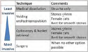
Methods of bladder urolith removal

References
1. ACVIM Small Animal Consensus Recommendations on the Treatment & Prevention of Uroliths in Dogs & Cats. https://onlinelibrary.wiley.com/doi/full/10.1111/jvim.14559.
2. Appel et al. Evaluation of risk factors associated with suture-nidus cystoliths in dogs and cats: 176 cases (1999–2006). J Am Vet Med Assoc. 2008.
3. Lulich J et al. (2013). Efficacy of two commercially available, low-magnesium, urine-acidifying dry foods for the dissolution of struvite uroliths in cats. J Am Vet Med Assoc. 243(8), 1147–1153.