K. Borgeat
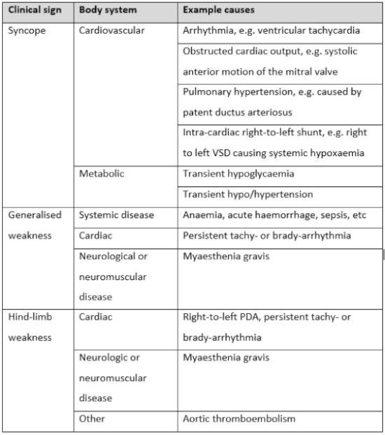
Syncope is defined as a “transient loss of consciousness and postural tone, associated with reduced cerebral delivery of oxygen and/or metabolites.” The commonest cause for syncope is cardiovascular (arrhythmias, outflow tract obstruction, etc.), but in rare cases it can be caused by transient hypo/hypertension or episodic hypoglycaemia.
Obtaining a detailed history, including the timeline of the episode and its association with recent events (exercise, respiratory signs, food intake, etc.), is absolutely crucial to choosing an appropriate diagnostic course for a cat presenting with syncope. If the clinician does not accurately identify the clinical signs—for example, if syncope is mistaken for a seizure episode—further testing may result in an incorrect diagnosis and treatment plan which influences patient well-being and further decision making over the coming months or years.
In dogs, syncope is generally rather easy to differentiate from a neurological or neuromuscular event. However, in cats, clinical signs perceived as neurological are often associated with a cardiogenic syncope event. In my experience, cats with bradyarrhythmias are commonly presented to the neurology referral service rather than a cardiologist in the first instance. History findings associated with syncope in dogs: association with exercise or exertion, falling over with flaccid muscles, unresponsive with eyes open, rapid recovery within 1–2 minutes. In cats, syncope is a more challenging event to identify so conclusively. It can occur at exertion or when lying still, is commonly associated with opisthotonos and limb rigidity, and occasionally is reported to feature vocalisation, salivation, facial motor activity or urination. These so-called “autonomic” signs are often suggestive to the clinician of a neurological event, and therefore heart disease could easily be overlooked. However, despite these clinical signs overlapping with neurological diseases, other features of syncope remain—namely the rapid recovery and return to normal activity. We will look at some video examples in the lectures.
One feline idiosyncrasy worth mentioning is that cats with bradyarrhythmias commonly present with facial motor activity alone, with no other signs of weakness or collapse. In this scenario, a diagnosis of a partial seizure is easily made. We would strongly recommend that any patient with “partial seizures” undergoes an ECG recording during an episode—whether this is lucky enough to be caught on a paper or digital ECG in-clinic, a Holter or an implantable loop recorder.
Diagnostic Investigation of Syncope
- History and video recording of episodes—vital.
- Physical examination: mucus membrane colour/CRT, arrhythmia, heart murmur, pulse quality/deficits, respiratory signs, behaviour and interactions in-clinic.
- Non-invasive blood pressure: Doppler systolic method.
- Blood tests: haematology, serum biochemical profile, cardiac biomarkers (NTproBNP and cardiac troponin I), endocrine testing (age dependent).
- ECG.
- Echocardiography—especially if murmur or arrhythmia.
- 24 h Holter ECG—next step to evaluate rhythm outside of the clinic, but poorly tolerated by most cats.
- Implantable loop recorder (Reveal device)—owner activates a subcutaneous ECG device to record during an episode of syncope.
| Figure 1. Paroxysmal asystole in a cat with episodic syncope | 
|
|
| |