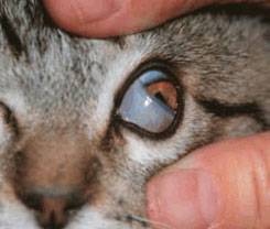
The normal third eyelid or nictating membrane sits in the inner corner of the eye. This shows a cat's normal third eyelid but the structure is basically the same in dogs. Photo by MarVistaVet
Prolapse of the Tear Gland of the Third Eyelid
Unlike humans (who only have two eyelids), dogs and cats have three. The third eyelid, technically called the nictitans or nictitating membrane, arises from the inner corner of the eye and covers the eye diagonally as shown. The eye is lubricated by tear film, which consists of water, oil, and mucus. The oil comes from glands lining the outer eyelids, the mucus comes from glands in the conjunctiva (the pink part inside the eyelids), and the water comes from tear (or lacrimal) glands. Each eye has two tear glands: one just above the eye and one located in the third eyelid. The gland in the third eyelid is believed to produce a full 30 percent of the total tear film water, so it is important to maintain the function of this gland.
The tear gland of the third eyelid is held in place by tissue fibers but some individuals have weaker fibers than they should so the gland protrudes. This protrusion is called a cherry eye. In the smaller breeds - especially Boston terriers, cocker spaniels, bulldogs, and beagles - the gland of the third eyelid is not strongly held in place for genetic reasons. The gland prolapses (drops down) out to where the owner notices it as a reddened mass. Out of its normal position, the gland does not circulate blood properly, may swell, and may not produce tears normally.
Treatment: Replacing the Gland in its Proper Location
By far the best treatment for cherry eye is replacing the gland back into its proper location. There are two techniques for doing this. The traditional tucking method (also called tacking) is probably the most commonly performed. Here, a single stitch is permanently placed, drawing the gland back where it belongs. Complications are uncommon but be aware of the following possibilities:
- If the stitch unties, the surface of the eye could become scratched by the suture. If this occurs, the eye will become suddenly painful and the suture thread may be visible. The suture can be removed and the problem solved.
- The tuck may not be anchored well enough to hold permanently. In fact, this surgery is notorious for this type of failure, and frequently a second or even third tuck is needed. If more than a couple of tucks have led to failure, it may be better to try the imbrication technique as described below. Some cases are repaired using both tuck and imbrication together.
- Sometimes cherry eye is accompanied by other eyelid problems that make the repair more difficult or less likely to succeed. In these cases, again, if the simple surgery is not adequate, ask your veterinarian if a referral to a veterinary ophthalmologist for the second surgery to maximize the chances of a permanent resolution is in the best interest of you and your pet.
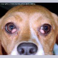
A dog with cherry eye in both eyes. Photo by Dr. Ian Herring
In a newer surgical technique called imbrication, or pocketing, a wedge of tissue is removed from directly over the actual gland. This technique is more challenging as it is not easy to determine how much tissue to remove. Tiny stitches that will eventually dissolve are used to close the gap so that the tightening of the incision margins pushes the gland back in place. Complications may include:
- Inflammation or swelling as the stitches dissolve.
- Inadequate tightening of the tissue gap may lead to recurrence of the cherry eye.
- Failure of the stitches to hold and associated discomfort. Loose stitches could injure the eye depending on the type of suture used.
Sometimes both surgical techniques are used in the same eye to achieve a good replacement. Harmful complications from cherry eye surgery are unusual but recurrence of the cherry eye can happen. If it recurs, it is important to let your veterinarian know so that a second surgery, either with your veterinarian or an ophthalmologist, can be planned.
Expect some postoperative swelling after cherry eye repair but this should resolve and the eye should be comfortable and normal in appearance after about a week. If the eye appears suddenly painful or unusual in appearance, have it rechecked as soon as possible.
Treatment: Removing the Gland
Historically, the prolapsed gland was treated like a small tumor; it was simply removed. This was before the full significance of the gland was realized.
If the third eyelid's tear gland is removed, it cannot be put back in place. If the other tear gland (the one above the eye) cannot supply adequate tears, not an uncommon phenomenon in older small breed dogs, then the eye becomes dry and uncomfortable. A thick yellow discharge results and the eye develops a blinding pigment covering for protection. This condition is called simply dry eye or more scientifically keratoconjunctivitis sicca and daily medical treatment is required to keep the eye both comfortable and visual. Not only is dry eye uncomfortable, but its treatment is often frustrating and time-consuming and there is expense involved. If left untreated, the eye can become blind. We would like the dog to maintain the greatest amount of tear-producing tissue possible, thus removing the gland for cosmetic reasons is not an acceptable treatment method.