Introduction
Reports related to the olfactory ventricle are scarce in the veterinary literature.
Objectives
The purpose of this study was to describe a dilation of the olfactory ventricle in an asymptomatic mature cat.
Methods
A 7-year-old mixed-breed female cat with no history of neurological signs was submitted to an MRI of the brain as part of a research study.
Results
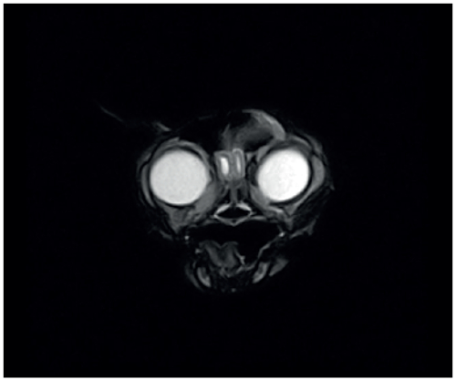
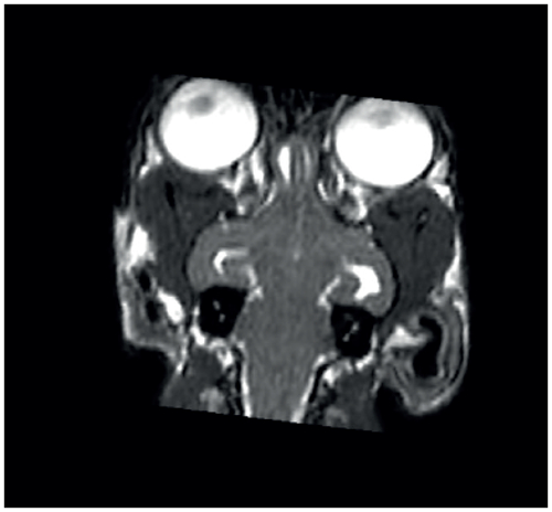
On MRI images, a cavity filled with cerebrospinal fluid was visualized in the central portion of the bilateral olfactory bulb. Both structures, which were compatible with olfactory ventricles since they communicated with the lateral ventricles, measured 7.60 mm in length. The left ventricle displayed a width of 1.30 mm and the right-sided ventricular structure exhibited a measurement of 2.60 mm. The height of the right and left olfactory ventricles were 5.70 mm and 2.00 mm, respectively. As well as in the dogs of a recent report, the enlargement of the olfactory ventricles occurred concomitant with lateral ventricular dilation. Unlike the asymptomatic cat of this study, the dogs presented neurological signs such as seizures. An increased intracranial pressure (ICP) - possibly related to the olfactory dysfunction - was assumed to be the producer of the neurological symptomology in these animals. In humans, symptoms like headache, nausea, vomiting and double vision have already been reported in subjects with ICP and olfactory disorder.
Conclusions
This study reports the olfactory ventricular enlargement in an asymptomatic mature cat.