Sandra M. May (Weltan), BVSc (Hons), MMedVet (KLD), PhD
Indications for Taking a Bone Marrow Sample
In general, bone marrow biopsy is a confirmatory or investigative tool used to follow-up on abnormal haematology results:
1. Nonregenerative anemia
2. Persistent thrombocytopaenia
3. Granulocytopaenia
4. Bi- or pancytopenia
5. Atypical cells in the peripheral blood (e.g., NRBC, blasts)
6. Staging lymphoma, mast cell and other tumors3
Contraindications:
1. Severe haemorrhagic diathesis
2. Thrombocytopaenia is not a contraindication
3. Infection is rare3
Protocol for Sampling
As with any laboratory test, sample quality is of the utmost importance if useful information is to be obtained. The following is a simple protocol for obtaining bone marrow samples:
Schedule Bone Marrow Aspiration and Cores Biopsies Early in the Day, if Possible
Bone marrow samples take longer to process, stain, and evaluate than other types of cytological or blood samples. Several slides may need to be examined, special stains may be done, and differential cell counts of 500–1000 cells are sometimes indicated.
Use Appropriate Analgesia
Bone marrow biopsies (both aspirates and cores) are painful. While many dogs require only local anesthesia, other dogs, cats and horses may need sedation or even general anesthesia. Plan for analgesia and sedation that will ensure adequate restraint for 20–35 minutes. Local anaesthetic should be injected under the skin and down to the periosteum overlying the injection site.2,4
Clip and Scrub the Site of Choice
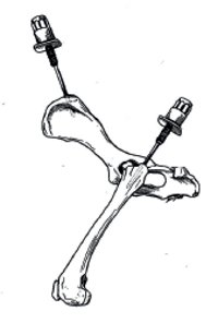
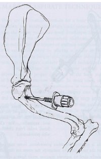
This may be the iliac crest, femur, humerus, or, in large animals, the sternum or ribs.3,4 The best site in birds is the proximal tibiotarsus.1
Use Appropriate Equipment
The following supplies and instruments are necessary:
 Bone marrow aspirate needle
 Bone marrow core biopsy needle
 Sterile 2% EDTA (add 0.36 ml sterile saline to a 4 ml EDTA tube and mix)
 12 cc syringe, 20 g needle, sterile gloves
 10% formalin for the core specimen
 Cytology and histopathology submission form
Obtain Both Aspirate and Core Specimens
A bone marrow aspirate is essential for evaluation of cell morphology. A core specimen is equally essential for evaluation of cellularity, stromal lesions, and the presence of focal lesions. A complete and accurate bone marrow evaluation cannot be made without both of these specimens or in the absence of a peripheral blood smear.
| Figure 1. Site for the iliac crest and proximal femur | 
From Grindem CB. Bone marrow biopsy and evaluation.Vet Clin North Am Small Anim Pract. 1989;19:669–696 with permission. |
|
| |
| Figure 2. Site for the proximal humerus | 
From Grindem CB. Bone marrow biopsy and evaluation. Vet Clin North Am Small Anim Pract. 1989;19:669–696 with permission. |
|
| |
Aspirate
1. With a 20 ga needle attached to the syringe, aspirate a small amount of EDTA; the amount of EDTA aspirated should fill the neck of the syringe, with just a drop in the syringe barrel, no more than 0.1 ml per 1.0 ml of bone marrow.
2. Remove the 20 ga needle and replace with a bone marrow aspirate needle.
3. Aspirate 0.5–1.0 ml of marrow. Remove syringe from needle and roll to mix.
4. Place marrow samples aspirated into the syringe into an EDTA tube from which excess EDTA has been shaken out.
5. Excessive EDTA (in syringe or tube) results in poor staining quality and an unreadable specimen.
Core Biopsy
1. Insert the biopsy needle through the same skin hole, but position it against the bone slightly anterior or posterior to the site of aspiration.
2. With the stylet in, firmly position the needle into the bone; remove the stylet and advance the needle to the depth or feel of the opposite cortex.
3. Using a twisting motion, loosen the core and remove the needle.
4. By inserting the stylet through the narrow end of the needle, push the sample into a small bottle of 10% formalin.
5. Make sure any sample being submitted for cytology (aspirates, blood) do not come in contact with fumes from open formalin containers.
Obtain a Sample for a Full Blood Count at the Time of Bone Marrow Biopsy
Interpretation
Normal progression:
|
Erythroid
|
Myeloid
|
Megakaryocytes
|
Monocytes
|
|
Rubriblast
|
Myeloblast
|
|
|
|
Prorubricyte
|
Promyelocyte
|
|
|
|
Rubricyte
|
Myelocyte
|
|
|
|
Metarubricyte
|
Metamyelocyte
|
|
|
|
Reticulocyte
|
Band cell
|
|
|
|
Erythrocyte
|
Mature granulocyte
|
|
|
Disease states:
1. Stem cell injuries:
a. Reversible:
i. Viral
ii. Drugs
iii. Chemicals
b. Irreversible:
i. Viral
ii. Idiopathic
2. Aplasia or hypoplasia:
a. Immune-mediated
b. Viral
c. Congenital?
3. Dysmyelopoiesis:
a. Immune-mediated
b. Drugs
c. Infectious diseases
d. Iron deficiency
e. toxicity
4. Myelodysplasia
5. Leukaemia
References
1. Campbell TW. Hematology of birds. In: Thrall MA, Baker DC, Campbell TW, De Nicola D, Fettman MJ, Lassen ED, Rebar A, Weiser G, eds. Veterinary Hematology and Clinical Chemistry. Oxford: Blackwell; 2006:225–258.
2. Harvey JW. Atlas of Veterinary Haematology: Blood and Bone Marrow of Domestic Animals. Philadelphia, PA: W.B. Saunders; 2001.
3. Moritz A, Bauer NB, Weiss DJ, Lanevschi A, Saad A. Evaluation of bone marrow. In: Weiss DJ, Wardrop KJ, eds. Schalm's Veterinary Hematology. Ames, IA: Wiley-Blackwell; 2010:1039–1046.
4. Thrall MA, Weiser G, Jain NC. Laboratory evaluation of bone marrow. In: Thrall MA, Baker DC, Campbell TW, De Nicola D, Fettman MJ, Lassen ED, Rebar A, Weiser G, eds. Veterinary Hematology and Clinical Chemistry. Oxford: Blackwell; 2006:149–178.