Histologic Features Associated with Spinal Deformity in Captive Sand Tiger Sharks
Abstract
This study was undertaken to investigate histologic features of the spinal deformity reported in captive Sand Tiger sharks (Carcharias taurus). Spinal segments from two adult Sand Tiger sharks with clinical signs of spinal deformity were compared to segments from a third Sand Tiger shark with a clinically normal spine. Distribution of mineralized tissue in affected and nonaffected spinal segments was determined by high-detail radiography. Unfixed, frozen tissue sections were examined with Nomarski interference microscopy and Raman microscopy. Formalin-fixed tissues were either embedded in methylmethacrylate, diamond saw sectioned and polished; or decalcified, followed by paraffin embedding and sectioning. Resulting sections were stained with toluidine blue, hematoxylin and eosin, or safranin O-fast green and then evaluated with standard transmitted or polarized light microscopy.
High-detail radiography revealed species-specific tesserae in the neural and hemal arches and the perichordal cartilage, and radiating patterns of mineralization in the centrum of the vertebrae from normal spinal segments (Figure 1). Prominent areas of ectopic mineralization occurred in the perichordal cartilage and connective tissue ventral to the vertebral centrum in the affected spinal segments (Figure 2).
| Figure 1 and Figure 2. | 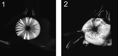
|
|
| |
Light microscopy of plastic embedded undecalcified sections and Nomarski interference microscopy of frozen tissue preparations revealed a prominent brown pigment in chondrocytes and the surrounding cartilage matrix from both non-affected and affected spinal segments. We could find no reference to this pigment in the literature regarding shark cartilage. Lack of previous documentation of the pigment may be related to our observation that the pigment was lost during standard histologic processing of frozen tissue preparations and during decalcification of fixed tissue.
In non-affected spinal segments, non-mineralized areas of the vertebrae contained the brown pigment (indicated by asterisk) in a defined lattice structure that appeared to be associated with distinct segments of fibrocartilage and an organized distribution of blood vessels from the perichondrium (Figure 3). The brown pigment was also seen in the mineralized areas of the vertebrae.
| Figure 3. | 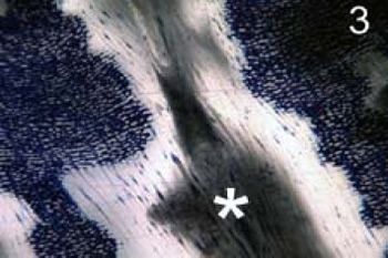
|
|
| |
In non-affected spinal segments, Raman microscopy was used to identify a characteristic calcium phosphate signal in the mineral from unfixed, frozen tissue preparations.
In affected spinal segments, areas of hyaline cartilage had numerous unique, pigment-mineral clusters. Nomarski interference microscopy indicated both angular and globular components to the clusters suggesting a potential protein-mineral composite (Figure 4). These clusters would increase to form large aggregates within the areas of ectopic mineralization (Figure 5) that were associated with disorganized vessels from the perichondrium.
| Figure 4 and Figure 5. | 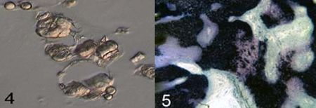
|
|
| |
Raman microscopy of the pigment-mineral clusters evaluated under standardized conditions revealed an attenuated calcium phosphate signal.
Morphologic evidence obtained in the present study suggests that the brown pigment discovered in Sand Tiger shark cartilage may be involved in either the organized recruitment of blood vessels or mineral deposition associated with normal growth of the vertebrae. The pigment mineral clusters characteristic of the affected spinal segments may reflect a modification of the pigment that is associated with the disruption of normal growth and the formation of ectopic mineralization. Future studies will be directed towards the isolation and identification of the brown pigment in normal and affected Sand Tiger shark cartilage in an attempt to discover its biochemical nature.
Acknowledgements
The authors appreciate the efforts of the husbandry staff from The Florida Aquarium, SeaWorld of Orlando, Aquarium of the Pacific and the Audubon Aquarium of the Americas for their efforts in the care of the animals and tissue collection used in this study.