Gregory K. Ogilvie, DVM, DACVIM (Internal Medicine, Oncology)
Adapted from: Ogilvie GK, Moore AS. Managing the Veterinary Cancer Patient. Trenton NJ, Veterinary Learning Systems. 1995, 542 pages. Ogilvie GK, Moore AS. Feline Oncology: A Comprehensive Guide for Compassionate Care. Trenton NJ, Veterinary Learning Systems. 2002, 550 pages. Ogilvie GK, Moore AS. Canine and feline Oncology: A Comprehensive Guide for Compassionate Care. Trenton NJ, Veterinary Learning Systems. 1-800-426-9119 vetlearn.com
Mast cell tumors present in such a wide variety of clinical signs: they are indeed the great impostors! They can look like anything and behave differently depending on the histologic type, location and the extent of the disease. The following is a brief discussion about these tumors. Some highlights are as follows:
 Mast cell tumor granules do not stain well with Diff Quick type stains unless they are "soaked" in the alcohol for several minutes prior to staining.
Some important prognostic indicators include duration of presence, location and histologic type in the dog.
Mast cell tumors tend to metastasize to nodes, liver spleen and bone marrow... rarely to lungs.
Radiation therapy is extremely effective for controlling local disease.
Prednisone and vincristine when used as single agents induce a remission (partial or complete) in about 23% of the tumors.
Vinblastine or CCNU with Prednisone is very helpful.
DIAGNOSIS OF MAST CELLS TUMORS
Diagnosis of mast cell tumors often can be made by a fine needle aspiration cytology but excisional biopsy is required if accurate histologic grading of the tumor is desired. Mast cell tumors are classified as round cell tumors along with lymphosarcoma, histiocytomas and transmissible venereal tumors.
Diagnostic workup of mast cells usually includes a number of procedures. These include a complete blood cell count (CBC), serum chemistry profile, and urinalysis. In addition, fine needle aspiration of the lesion, regional lymph nodes and examination of buffy coats or bone marrow helps to determine the extent of tumor involvement. A CBC is valuable in assessing animals with mast cell tumors because those animal patients with systemic mastocytosis occasionally have peripheral eosinophilia and basophilia in addition to circulating mast cells. Mastocytemia is a more common clinical phenomenon in the cat than in the dog. The CBC may also give evidence of gastrointestinal bleeding or gastrointestinal perforation. In general, mastocytosis associated with primary cutaneous tumors is more easily detected by examination of the buffy coat or bone marrow than by examination of peripheral blood. Care must be exercised in interpreting buffy coats since mastocytemia has been reported in a variety of acute inflammatory diseases of the dog including parvovirus infections. Peripheral mast cell counts may be high in cats with mastocytosis and have accounted for up to 25% of the total white cell count.
| Figure 1. | 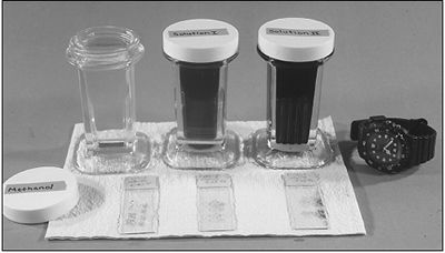
Mast cell tumors generally do not stain well with Diff Quick Stain unless the slide is placed in the methanol for at least 2 minutes prior to regular staining. |
|
| |
Buffy Coat Smears and Bone Marrow Aspirate
Buffy coat smears of blood samples may be examined microscopically for the presence of mast cells but bone marrow smears appear to be more sensitive and are not associated with as many false positives. Bone marrow evaluations should be performed in animals with mast cell tumors. Recent studies have demonstrated that normal dogs have less than 1 mast cell per 1,000 cells in the bone marrow. Veterinary investigators suggest mast cells in greater concentrations than 10/1,000 cells is abnormal.
Lymph Node Aspiration
Any animal patient with mast cell tumors should be carefully examined for lymphadenopathy in areas draining the primary tumor. Enlarged lymph nodes should be examined for the presence of mast cells as evidence of tumor spread. Such findings have important implications with regard to therapeutic strategies.
Radiology
Abdominal radiographs may be useful in evaluating dogs and cats. This is especially true in cats because of the high incidence of splenic involvement in this species with mast cell tumors. Chest radiographs rarely identify the presence of pulmonary metastases. In cases of mast cell tumors that involve the hind-limbs, perineal or preputial area, abdominal radiographs may be helpful in detecting metastatic lymphadenopathy in the iliac and sublumbar lymph nodes.
Miscellaneous
Tests Occult blood tests may be useful in evaluating patients with mast cell disease. The stools of dogs with mast cell tumors should be examined for the presence of gastrointestinal bleeding as evidence of GI ulceration. In many cases, feces may contain small amounts of blood that are insufficient to produce melena. Gastrointestinal bleeding can be identified by chemical tests based on blood peroxidase activity that involves catalyzing the conversion of hydrogen peroxide to water and oxygen. The most sensitive test contained orthotoluidine or benzidine as a chemical oxidizer to a color product. These tests are so sensitive that false positives may result if the diet has contained red meat for up to three days before testing. Therefore, careful examination of GI bleeding should be made in mast cell cases and indeed, many patients are routinely treated to block the effects of mast cell hyperhistaminemia or that results in increased gastric acid secretion in GI ulceration.
THERAPY
Surgical considerations include wide surgical margins with at least 3 cm of normal looking skin around the tumor should be removed when possible. The 3 cm recommendation is a guideline and might not be feasible when the tumor is located on the face, lower limbs or in the inguinal region. It should be remember-ed that most mast cells extend laterally to adjacent tissue rather than deep into underlying muscles. All excised tumor should be examined histologically for the completeness of excision. Extension of the tumor beyond the surgical borders should prompt either wider excision or radiation therapy of the tumor bed. Approximately 50% of the mast cell tumors recur at the surgical site traditionally. Histologic grade is an important factor in predicting recurrence at the surgical site. Those that are undifferentiated tend to have a higher recurrence rate. Cats with mast cell tumors with splenic involvement often will benefit from splenectomy. Survival times of 10 weeks to 30 months have been reported following splenectomy, even in patients with evidence of systemic mastocytosis.
Seguin et al (J Am Vet Med Assoc 218[7]:1120-1123 2001) evaluated 60 mast cell tumors that were surgically excised with clean margins in 55 dogs were included. Median follow-up time was 540 days. Three mast cell tumors recurred locally; median time to local recurrence was 62 days. Six dogs developed another mast cell tumor at a different cutaneous location; median time to a different location was 240 days. Three dogs developed metastases; median time to metastasis was 158 days. The authors concluded that additional local treatment may not be required after complete excision of grade-II mast cell tumors and that most dogs do not require systemic treatment.
Glucocorticoid therapy frequently results in partial or occasionally complete remissions in canine mast cell tumors. However, cats appear to be less responsive to glucocorticoid treatment. The effect of glucocorticoids is to reduce markedly the number of mast cells in the mast cell tumor. The exact mechanism by which glucocorticoids exert their cytotoxic effects on mast cell tumors is unknown although it may be similar to the effects of glucocorticoids on lymphocytes. The susceptibility of mast cell tumors might depend on the presence of intracytoplasmic glucocorticoid receptor sites. Glucocorticoid receptor sites have recently been found in the cytoplasm of canine mast cell tumors. Although sex steroid receptors for progesterone and estrogen have been recently described in dogs with canine mast cell tumors, the role of sex steroids in the treatment of canine mast cell tumors has yet to be investigated. The type of glucocorticoids administered appears to be unimportant but it has been suggested that intralesional corticosteroid may be more effective than systemic therapy for local disease. Fewer Cushingoid side effects have been seen with short-acting glucocorticoids such as prednisone or prednisolone when used in the dog. The usual dose of prednisone is .5 mg/kg orally administered once daily and that of triamcinolone is 1 mg for every cm diameter of tumor intralesionally, administered every two weeks. Remission times are usually 10 to 20 weeks. Dogs that are tumor free after six months however have a low incidence of recurrence and therefore therapy is usually discontinued at this time. Tumor resistance may be caused by the emergence of mast cells with fewer or ineffective glucocorticoid receptors. Survival data based on histologic grade correlates with various chemotherapeutic regimens has not been reported.
Vinblastine and prednisone or CCNU appear to be the most favored drug protocols for the treatment of mast cell tumors. The use of these drugs is always with surgery.
Rassnick and colleagues (J Vet Intern Med 13[6]:601-605 1999) evaluated the efficacy and toxicity of CCNU in 23 dogs with measurable mast cell tumors (MCT). Response could be evaluated in 19 dogs. Eight of the 19 dogs (42%) had a measurable response to CCNU. One dog had a durable complete response for 440 days. Seven dogs had a partial response for a median and mean duration of 77 days and 109 days, respectively (range, 21-254 days). The acute dose-limiting toxicity was neutropenia 7 days after administration of CCNU.
Thamm et al (J Vet Intern Med 13[5]:491-497 1999) evaluated 41 dogs with mast cell tumors treated with oral prednisone and vinblastine both in the adjuvant setting and in dogs with gross disease. Adverse effects were noted in 20% of the patients, usually after the 1st dosage. Median survival time (MST) for the entire patient population was not reached with a median follow-up of 573 days; however, the MST for dogs with grade 111 MCT was 331 days, with 45% of dogs alive at 1 and 2 years.
Ancillary drug therapy is important with canine mast cells. Animals with mastocytosis or palpable mast cell disease should receive H2 antagonists. Cimetidine (Tagamet) reduced gastric acid reduction by competitive inhibition of the action of histamine on H2 receptors of the gastric parietal cells. Ranitidine (Zantac, Glaxo Inc, Fort Lauderdale, FL), a newer H2 antagonist that requires less frequent ad-ministration, is in some clinics. The objective of the therapy is to prevent gastro-intestinal ulceration associated with elevated levels of histamine and to treat ulcers already present. Some new evidence indicates that cimetidine may also alter the immune response to this tumor as well as activation of certain alkylating agents. Dogs and cats with evidence of gastrointestinal ulceration and bleeding might also benefit from sucralfate (Carafate, Marion Labs Inc, Kansas City, MO) therapy. Sucralfate reacts with stomach acid to form a highly condensed viscous adherent paste-like substance that binds to the surface of both gastric and duodenal ulcer sites. The barrier formed at the ulcer site protects the ulcer from potential ulcerogenic properties of pepsin, acid and bile allowing the ulcer to heal. Because sucralfate interferes with absorption of cimetidine, these two drugs should be given at least two hours apart. The usual dosage of sucralfate is 1 gm given orally. H1 antagonists such as Benadryl should be used along with cimetidine prior to and following surgical removal of canine mast cell tumors to help prevent the negative effects of local histamine release on fibroplasia wound healing. H1 antagonists also should be used with cryosurgery or hyperthermia therapy. Another re-commended ancillary medication is an antiserotonin agent (cyproheptadine). The use of this drug is controversial since serotonin has only been identified in rat and mouse mast cells and definitive studies in the dog and cat are lacking. The use of drugs that stabilize mast cells (sodium cromoglycate) have been described in the treatment of human patients with mastocytosis but not in animals.
Radiotherapy has been used alone or in combination with other treatment modalities. Most reports indicate remission rates of 48 to 77%. Doses of 3,000 to 4,000 rads were used in these studies. Total radiation therapy is usually fractionated and delivered over a period of three to four weeks. The use of radiotherapy is somewhat expensive and is confined to referral centers. Mast cell tumors in regional lymph nodes and bone marrow appear to be more resistant to the effects of radiotherapy than those confined to the skin. Response of mast cell tumors to radiation therapy may correlate to histologic grade but has not been studied.
PROGNOSIS
The natural behavior of mast cells suggests prognosis of this tumor depends on the species, breed, histologic grade, tumor location, clinical stage and growth rate. The higher the histologic grade (more undifferentiated tumor), the poorer the prognosis. This criteria has not had universal acceptance however, probably due to the precise nature of histologic grading as well as tumor heterogeneity. Clinical staging and the extensive-ness of microscopic tumor masses beyond what might be detected clinically also plays an important role in the failure of universal acceptance of the histologic grading system Tumor location is considered by many investigators to be an important prognostic feature Clinical stage is a clinical means of assessing tumor spread of the disease process. The higher the clinical stage, the more guarded the prognosis. A high histologic grade, however, should increase the clinical stage at least one level. Growth rate but not tumor size is determined also to be an important prognostic indicator. Growth rate reported by Bostock indicates that dogs that have tumors that grow greater than 1 cm per week have only a 25% chance of living an additional 30 weeks.
References
1. Ogilvie GK, Moore AS. Mast Cell Tumors. In: Managing the Veterinary Cancer Patient: A Practice Manual. Trenton: Veterinary Learning Systems. 1995:503-514.
2. Rassnick KM, Moore AS, Williams LE, London CA, et al. Treatment of Canine Mast Cell Tumors with CCNU (Lomustine) J Vet Intern Med 13[6]:601-605 1999.
3. Thamm DH, Mauldin EA, Vail DM. Prednisone and Vinblastine Chemotherapy for Canine Mast Cell Tumor-41 Cases (1992-1997) J Vet Intern Med 13[5]:491-497 1999
4. Seguin B, Leibman NF, Bregazzi VS, et al. Clinical Outcome of Dogs with Grade-II Mast Cell Tumors Treated with Surgery Alone: 55 Cases (1996-1999). J Am Vet Med Assoc 218[7]:1120-1123 2001.