Sean M. Perry1*; Mary Beth Tims1; Nicole I. Stacy2; Jennifer Dill-Okubo3; Jill Arnold4; Taya Marquardt Ezell5; Alexa J Delaune1
Abstract
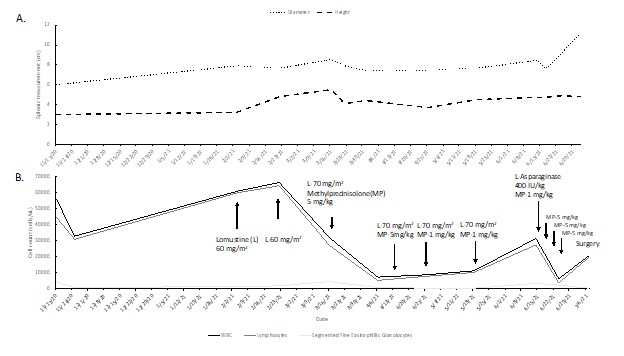
Neoplasia in elasmobranchs is rarely reported and management of clinical neoplasia in elasmobranchs has not been reported in the literature. Prevalence of neoplasia in elasmobranchs under human care is reported to be 0.5–4% based on two retrospective studies evaluating elasmobranch cases submitted for pathology.1,2 In batoids, disseminated lymphoid neoplasia and splenic round cell tumors have been described.1,3 There are no published reports of chemotherapeutic interventions for lymphoid neoplasia in elasmobranchs. A two-year-old cownose ray (Rhinoptera bonasus) (CNR) was evaluated for annual examination. Coelomic ultrasound showed hepatomegaly with a hyperechoic echotexture and an enlarged spleen measuring 6 cm wide x 3 cm height at the largest point with mixed echogenicity. Enlarged hepatic vessels were observed in the left caudal liver lobe. Bloodwork revealed anemia and a leukocytosis with immature lymphocytes within the peripheral blood. An additional diagnostic workup was performed including ultrasound guided fine needle aspirate (FNA) and coelioscopy under general anesthesia. Splenic and liver biopsies were obtained. FNAs of the spleen revealed reactive lymphoid hyperplasia and extramedullary hematopoiesis. Hepatic aspirates showed heterogenous lymphoid infiltrate, hepatocellular degeneration, and hyperplasia. Splenic tissue was not recovered with endoscopy; however, hepatic biopsies showed lymphoma. Following diagnosis, chemotherapy was initiated. Due to species specific concerns in successful administration of chemotherapy, a protocol consisting of oral lomustine (60–70 mg/m2) and injectable methylprednisolone (1–5 mg/kg) was initially employed every 3 weeks. Following administration of lomustine staff did not service the holding pool or associated life support for three days. To monitor response to therapy serial complete blood counts, biochemistries, and coelomic ultrasounds were performed. Image 1A illustrates the changes in splenic size throughout therapy. Image 1B documents the chemotherapy protocol used to treat multicentric lymphoma and the changes observed in the white blood cells, lymphocytes, and segmented eosinophilic granulocytes during therapy. Treatment was performed for 6 doses of lomustine (4 months). The most observed changes in splenic size and white blood cell counts were associated with administration of methylprednisolone. Following the initial course of treatment, the animal demonstrated signs of being refractory to therapy therefore L-Asparaginase (400 IU/kg) was administered as a rescue therapy in addition to increased administration of methylprednisolone. Despite these medications, splenic size increased. Due to a declining appetite presumably secondary to a mass effect, surgical intervention was elected to improve quality of life. A splenectomy was performed under general anesthesia; following successful splenic removal, hemostasis could not be maintained without compromising the gastrointestinal vasculature. The animal was humanely euthanized intraoperatively. A full necropsy was performed and histopathology showed lymphoma present within the liver, spleen, inter-renal organ, epigonal and Leydig organ. All immunohistochemistry staining was negative. Methylprednisolone (1–5 mg/kg) shows potential as an effective therapeutic to treat lymphoid neoplasia in elasmobranchs. Lomustine and L-Asparaginase may be have some synergistic effect with methylprednisolone in treating lymphoma in elasmobranchs; however, further clinical cases or research is needed to demonstrate efficacy and determine proper dosing regimens.
Figure 1

A. Ultrasonographic splenic measurements during treatment for multicentric lymphoma.
B. Chemotherapy protocol and total cell counts during treatment. Arrows indicate a point in time the animal was treated with chemotherapy. Each arrow has the drug name and respective dose.
Acknowledgements
We thank the animal and facilities staff at the Mississippi Aquarium for their care of this animal.
Literature Cited
1. Garner MM. 2013. A retrospective study of disease in elasmobranchs. Veterinary Pathology, 50(3), 377–389.
2. Stidworthy MF, Thorton SM, James R. 2017. A review of pathologic findings in elasmobranchs: A retrospective case series. In: Smith M, Warmolts D, Thoney D, Hueter R, Murray M, Ezcurra J, eds. Elasmobranch husbandry manual II: Recent advances in the care of sharks, rays, and their relatives. Columbus (OH): Ohio Biological Society. p 277–288.
3. Stilwell JM, Camus AC, Zachariah TT, McManamon R. 2019. Disseminated lymphoid neoplasia and hepatoblastoma in an Atlantic stingray, Hypanus sabinus (Lesueur 1824). J Fish Dis. 42: 319– 323.