Total Ear Canal Ablation and Lateral Bulla Osteotomy (TECA-LBO) for Successful Surgical Management of Otitis Media in Harbor Seals (Phoca vitulina)
1College of Veterinary Medicine, Purdue University, West Lafayette, IN, USA; 2Mystic Aquarium, Mystic, CT, USA; 3Metropolitan Veterinary Hospital, Akron, OH, USA; 4Cape Cod Veterinary Specialists, Buzzards Bay, MA, USA; 5National Marine Life Center, Buzzards Bay, MA, USA
Abstract
Chronic, severe otitis media was diagnosed in four Atlantic harbor seals (Phoca vitulina concolor), three of which were stranded animals undergoing rehabilitation. All seals presented with unilateral purulent aural discharge that intermittently recurred despite prolonged topical and systemic antimicrobial therapy. Aerobic culture from aural exudates isolated multi-drug resistant organisms in all seals, including Pseudomonas aeruginosa, Staphylococcus pseudintermedius, Klebsiella pneumoniae, and/or Enterococcus faecalis. Computed tomography (CT) was performed in three cases to confirm otitis media and positive contrast ear canalography was used in one case to confirm tympanic membrane rupture. Given the persistent nature of otitis, surgical intervention was indicated. Presurgical planning included dissection of postmortem seal specimens and review of museum specimens to create an anatomic rendition of important neurovascular anatomy relevant to performing a total ear canal ablation and lateral bulla osteotomy (TECA-LBO) (Figure 1). TECA-LBO was successful in achieving complete clinical resolution of otitis in all seals. Postoperative complications included temporary unilateral paralysis of the left nare in two cases and a transient left ptosis in one animal. Partial to complete surgical site dehiscence occurred in all cases but complete healing was achieved by second intention in 60 days or less. One rehabilitated seal was fitted with a satellite tag that confirmed normal swimming and diving patterns post-release. In harbor seals, TECA-LBO can be performed safely to treat persistent cases of otitis media and should be considered in cases of chronic otitis that are not responsive to medical management.
Figure 1
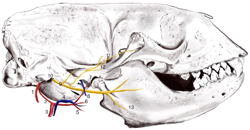
Medical illustration depicting the anatomical location of important neurovascular structures in the Atlantic harbor seal (Phoca vitulina concolor) relevant to performing a total ear canal ablation and lateral bulla osteotomy (TECA-LBO). These structures include the (1) internal carotid artery, (2) caudal auricular artery, (3) external carotid artery, (4) maxillary vein, (5) lingual artery, (6) facial artery, (7) maxillary artery, (8) retroarticular vein, (9) facial nerve (CN VII), (10) stylomastoid foramen, (11) retroauricular foramen, (12) auriculotemporal nerve (branch of CN V), and (13) lingual nerve.
Acknowledgements
The authors thank Molly E. Bryant for the medical illustration, Dr. Allison Tuttle for her guidance and mentorship, Drs. James Bailey and Louisa Rahilly for their anesthesia support, and the past and present husbandry staff and volunteers at Mystic Aquarium and National Marine Life Center.
*Presenting author
+Student presenter