Pregestational, Gestational, and Postgestational Serum Progesterone, β-Human Chorionic Gonadotropin, and Estradiol Levels in a Bornean Orangutan (Pongo pygmaeus pygmaeus)
Abstract
Urinary hormonal values of estrogen, pregnanediol, estrone, estradiol and androsterone during pregnancy and menstrual cycles of orangutans have been studied by several investigators.4-7,9 Serum values for estradiol, progesterone, testosterone and luteinizing hormone (LH) have been evaluated during the menstrual cycles of orangutans and macaques.10,12
In this study, serum estradiol-17β, progesterone, and β-human chorionic gonadotropin (β-HCG) were monitored before, during and after a pregnancy in a 22-yr-old female Bornean orangutan (Pongo pygmaeus pygmaeus). Results were compared to a non-pregnant 33-yr-old female Bornean orangutan in the same facility and human reference normals. Blood samples were obtained during weekly to biweekly conditioning programs without sedatives or tranquilizers. Trans-abdominal ultrasound images were obtained during the later stages of pregnancy.
Serum progesterone (4-prenen-3,20-dione), β-HCG, and estradiol [1,3,5 (10)-estratrien-3,17 β-diol] values were determined using microparticle enzyme immunoassays (AxSYM®, Abbott Laboratories, Abbott Park, Illinois 60064 USA) performed on samples obtained from the orangutan through a stationary conditioning program. These samples were frozen and analyzed at a later date.
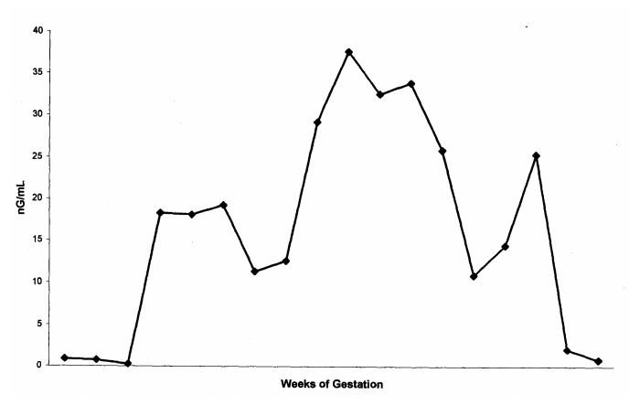
Values for serum progesterone, β-HCG, and estradiol are presented in Figs. 1–3. Results of pregestational, gestational and post-gestational values closely parallel human normals.2,13,14 In humans progesterone is produced primarily by the corpus luteum of the ovary.1 Progesterone prepares the uterus for implantation and maintains the uterus during pregnancy. During the menstrual cycle, serum progesterone values elevate to 10–20 ng/ml 5–7 days after ovulation. During the 6th wk of pregnancy, the placenta becomes the major source of progesterone.8 Values during this time elevate from 10–50 ng/ml during the first trimester to 50–2,809 ng/ml during the third trimester.2,13 In this case, progesterone levels were between 10–15 ng/ml during the first trimester, and elevated to 30–40 ng/ml during the second trimester. However, progesterone levels decreased to levels of 10–15 ng/ml during the third trimester (Fig. 1). Serum levels began to elevate to previous levels, but never fully recovered. Reasons for this are unknown. Although this pregnancy proceeded to term, the infant was stillborn. An underlying cause was not determined.
Figure 1. Serum progesterone values (ng/ml)
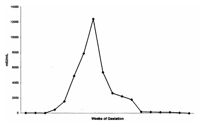
β-HCG appears to maintain the corpus luteum during early pregnancy in humans, allowing continued secretion of progesterone, which supports the endometrium. Values for serum β-HCG in humans demonstrate a maximal peak in the late first trimester, with a decline to a fairly constant level by mid gestation.2 In our study, a similar peak and return to lower, constant levels was observed (Fig. 2).
Figure 2. Serum Beta-human chorionic gonadotropin values (mIU/ml)
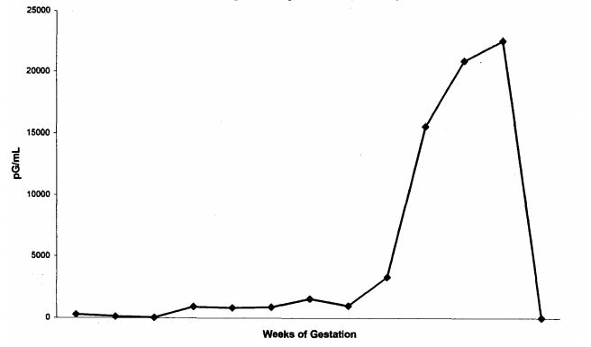
Estradiol levels in human studies demonstrate a constant rise until parturition.11,14 Levels of 100–500 pg/mL occur during the first trimester, 5,000–15,000 during the second trimester, and 10,000–40,000 pg/mL during the third trimester.14 A similar curve was seen in this group of data for the orangutan (Fig. 3).
Figure 3. Serum estradiol values (pg/ml)
To our knowledge, this is the first report of serial serum levels of progesterone, β-HCG, and estradiol before, during and after gestation in an orangutan. A similar sampling is being obtained in the same animal during its current gestation. Results are being correlated with weekly trans-abdominal ultrasound and daily urinary levels of progesterone, β-HCG, and estrogen. It is hoped that these values can be used to provide a basis for comparing the reproductive hormones of orangutans during pregnancy.
Acknowledgments
The authors would like to thank the orangutan keeper staff, which has worked with the veterinary staff for the past 6 yr perfecting the conditioning programs with the orangutans at the Kansas City Zoological Gardens.
Literature Cited
1. Abraham G.E., W.D. Odell, 1972. Simultaneous radioimmunoassay of plasma FSH, LH, progesterone, 17-hydroxyprogesterone, and estradiol-17β during the menstrual cycle J. Clin. Endocr. 34:312–318.
2. Braunstein G.D., J. Rasor, D. Adler, H. Danzer, and M.E. Wade. 1976. Serum human chorionic gonadotropin in normal and pathologic pregnancy. J. Clin. Endocrinol. Metab. 126:678–681.
3. Buster, J.E. and G.E. Abraham. 1975. The application of steroid hormone radioimmunoassays to clinical obstectrics. Obstet. Gynecol. 46(4):489–499.
4. Collins, D.C., C.E. Graham, and J.R. K. Preedy. 1975. Identification and measurement of urinary estrone, estradiol-17β, estriol, pregnanediol and androsterone during the menstrual cycle of the orangutan. Endo. 96(1)93–101.
5. Czekala N.M., K. Benirschke, H. McClure, and B.L. Lasley. 1983. Urinary estrogen excretion during pregnancy in the gorilla (Gorilla gorilla), orangutan (Pongo pygmaeus) and the human (Homo sapiens). Biol of Repro. 28:289–294.
6. DeGroot L.J. (ed.) Endocrinology. 2nd edition, vol 3, Ovarian Hormone Synthesis, Circulation, and Mechanism of Action. W.B. Saunders Co, Philadelphia, 1989.
7. Graham C.E. 1988. Reproductive Physiology. In: Schwartz J.H. (ed.) Orangutan Biology. Oxford University Press, New York. 91–101.
8. Hertig, A.T. and R.B. Livingston. 1944. Spontaneous, threatened and habitual abortion: their pathogenesis and treatment. N. Eng. J. Med. 230:797–805.
9. Lasley, B.L., J.K. Hodges, and N.M. Czekala. 1980. Monitoring the female reproductive cycle of great apes and other primate species by determination of oestrogen and LH in small volumes of urine. J. Reprod. Fert. 28:121–129.
10. Nadler R.D., D.C. Collins, and M.S. Blank. 1984. Lutenizing hormone and gonadal steroid levels during the menstrual cycle of orangutans. J. Med. Primatol 3:305–314.
11. Ryan, K.J. 1980. Placental synthesis of steroid hormones. In: Tulchinsky D, K.J. Ryan (eds.) Maternal-fetal Endocrinology. W.B. Saunders Co. 3–16.
12. Shideler S.E., C.J. Munro, L. Tell, G. Owiti, L. Laughlin, R. Chatterton, and B.L. Lasley. 1990. The relationship of serum estradiol and progesterone concentrations to the enzyme immunoassay measurements of urinary estrone conjugates and immunoreactive prenanediol-3-glucuronide in Macaca mulatta. Am. J. Primatol. 22:113–122.
13. Speroff, L., R.H. Glass, and N.G. Kase. 1994. The Endocrinology of Pregnancy. In: Mitchell, C. (ed.). Clinical Gynecologic Endocrinology and Infertility, 5th edition. Williams and Wilkins. 251–289.
14. Winkel, P., P. Gaede, and J. Lyngbye. 1976. Method for monitoring plasma progesterone concentrations in pregnancy. Clin. Chem, 22:422–428.