Kathryn C. Gamble1, DVM, MS, DACZM; Michael M. Garner2, DVM, DACVP; James T. Raymond2, DVM, DACVP; Laura Krause1, RVT; Thomas P. Alvarado1, DVM, MS
Abstract
From 1990 to 2000, 10 cases of pancreatic islet fibrosis were documented in the Dallas Zoo collection of 64 rock hyrax (Procavia capensis). Eight (one male, seven females) of these animals entered the collection as adults and were presumed to be unrelated to each other. Four of the females arrived in 1993 pregnant by wild, unknown males, and produced nine (two males, four females, three unsexed) offspring, collectively. In 1994 and 1995, the single captive adult male sired 47 (17 males, 26 females, 4 unsexed) offspring from these wild-caught females and one of their 1993 offspring. During the late 1990s three rock hyrax presented with clinical diabetes mellitus (Cases 4, 6, and 10), ultimately, related to pancreatic islet fibrosis. Additional cases of pancreatic islet fibrosis were documented in four unrelated (Cases 1, 2, 3, and 5) and five related animals (Cases 6–10). This clustering of disease supported a review of the medical history, family tree, and pathology for this species in the collection (Figure 1, Table 1).
Figure 1
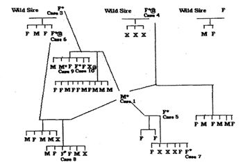
Pedigree of rock hyrax (Procavia capensis) at the Dallas Zoo (1990–2000) with presentation of pancreatic islet fibrosis. M=male, F=female, U=stillborn; *pancreatic islet fibrosis (post-mortem); @=clinical diabetes mellitus (ante-mortem). Note: Case 2 is not related to the family tree presented. Case 5 is a wild-caught female from 1993 that was not pregnant by a wild sire.
Table 1. Signalment, medical history, and pathology of rock hyrax (Procavia capensis) at the Dallas Zoo affected with pancreatic islet fibrosis (1990–2000)
|
Case number
|
Age
|
Gender
|
Clinical signs
|
Islet fibrosis
|
Other pathology
|
|
1
|
6 y
|
M
|
Wasted body condition
dental disease
azotemia
|
3+
|
Nephrosclerosis
gastric ulceration
cystitis
|
|
2
|
2 y
|
F
|
Comatose
pneumonia
|
4+
|
Purulent pneumonia
|
|
3
|
∼4 y
|
F
|
Comatose
alopecia
wasted body condition
|
4+
|
Cardiomyopathy
gastritis
hemosiderosis
nephritis
|
|
4
|
∼6 y
|
F
|
Euthanasia
weakness
azotemia
diabetes mellitus
|
3+
|
Hemosiderosis
glomerulosclerosis
|
|
5
|
∼3 y
|
F
|
Found dead
|
3+
|
Myeloproliferative disease
hemosiderosis
myocardial hypertrophy
|
|
6
|
7 y
|
F
|
Euthanasia
diabetes mellitus
|
4+
|
Hemosiderosis
glomerulosclerosis
lingual candidiasis
|
|
7
|
1 y
|
F
|
Found dead
|
3+
|
Lymphoma
|
|
8
|
1 y
|
F
|
Comatose
|
3+
|
Nephritis
sepsis
|
|
9
|
4 y
|
M
|
Chronic diarrhea
found dead
|
4+
|
Hemosiderosis
glomerulosclerosis
|
|
10
|
4 y
|
F
|
Euthanasia
diabetes mellitus
|
4+
|
Hemosiderosis
cardiomyopathy
glomerulosclerosis
|
Both the Dallas Zoo and five other institutions maintained offspring from this line. Review of the medical history provided minimal insight to the disease etiology. Clinical signs were vague and insidious in onset: rough hair coat, soft but not unformed feces, and weight loss. Minor trends in clinical pathology were identified in those animals with islet fibrosis. All but one animal (Case 6) presented with persistent hyperglycemia. All animals presented with persistent elevation in creatinine phosphokinase and amylase. Lipase concentrations were steady (9–39 U/L) until shortly before presentation of clinical signs. Although ISIS data were consulted for amylase and lipase, it is likely that animals in this line were providing data to these physiologic reference ranges.
Histologically, lesions were similar in all affected animals. The pancreas had severe islet fibrosis with extension of the fibrous tissue into the surrounding interstitium and associated disruption of the acinar structures. Density of islet cells in the areas of fibrosis was variable, with some islets having few or no islets cells but others having approximately normal cellular density. Cases with mild fibrosis had islet cell hyperplasia. Occasionally, ducts were mildly dilated and contained inspissated secretory material. A battery of special stains was performed on the pancreas from Case 7. Iron and copper stains were negative. Sclerotic sections of the pancreas were negative for amyloid with Congo red. A trichrome stain confirmed that the islet deposition was collagen and accentuated that distribution was earliest and most severe around and in the islets. A Churukian-Schenk stain for endocrine granules accentuated the presence of islet cells in areas of early interstitial fibrosis, further supporting the islet as the initial site of fibrosis. Electron microscopy performed on Case 7 supported the histologic findings and revealed extensive collagen deposition in the interstitium between clusters of sequestered islet and acinar cells. Frequent fibroblasts were associated with the collagen deposition. Islet cells were partially degranulated, and some had vacuolar changes in the nucleus. No viral agents or inflammatory processes were detected. The cause for this condition could not be determined morphologically. Additional significant lesions in many of these animals included hemosiderosis, glomerulonephritis or glomerulosclerosis, opportunistic infections, and degenerative cardiac disease; all of which are common complications of diabetes in mammals.
Differential diagnoses for this novel pancreatic lesion in adult rock hyrax include nutritional toxicity, infectious source, and genetic predisposition that manifests with age. Diet review, clinical pathology, and evaluation of serum for autoantibodies to the pancreatic islets will be pursued in this species.
Acknowledgments
We appreciate those institutions responding to our queries: Caldwell Zoo, Denver Zoological Gardens, Staten Island Zoo, Cleveland Metroparks Zoo, and Chehaw Wild Animal Park. Consultation on the family tree for genetic transmission and pancreatic autoantibody testing was provided by Dr. Helen Hobbs of the University of Texas Southwestern Medical School, Dallas, Texas, and Dr. Ake Lernmark of the University of Washington, Department of Medicine, Seattle, Washington. Technical support was provided by Roy Brown at Histology Consulting Service, Oak Harbor, Washington.