S. Boysen
Faculty of Veterinary Medicine, Veterinary Clinical and Diagnostic Sciences, University of Calgary, Calgary, AB, Canada
Introduction
Emergency venous cutdowns are a rapid, non-sterile technique used to surgially expose superficial peripheral veins for rapid visual IV catheter placement. Indications include patients presenting in shock where IV access is hampered or patients in which multiple percutaneous IV catheter attempts have failed (often defined as more than 5 minutes to place a peripheral catheter and/or more than 3 attempts to pass a percutaneous IV catheter). If vascular access is not achieved with these guidelines, an emergency venous cutdown should be attempted. With practice (cadavers are good models to practice), most clinicians, can perform this procedure faster and more reliably than placing an intraosseous catheter manually. Newer automated IO techniques (EZIO) however are faster than cut downs. In puppies or kittens with small vessels, the manual IO route is preferred because the cortex of the bone is soft in these patients and the IO space easily accessed with manual IO catheters. In more stable cardiovascular patients, ultrasound guided IV catheters can be placed into peripheral veins.
Any peripheral vein can be used for a cut down, including the saphenous, jugular and cephalic, however, the saphenous is often used in dogs, while the medial cutaneous saphenous vein (using a smaller 22- to 25-ga catheter is often used). If peripheral veins are difficult to find a jugular cutdown can be used in either cats or dogs.
Given the contaminated nature of cutdowns, once the patient is resuscitated and a sterile catheter can be place the cutdown catheter should be removed. Liberal flushing of the incision site with the catheter in place, before removal, is recommended. If the vein bleeds continuously following removal (rare in the absence of coagulopathies) the vessel can be ligated. The wound should not be fully closed to allow drainage and can be covered with a bandage for 48 hours, checking the incision and changing the bandage as needed.
Equipment
- Clippers
- Surgical scrub solution
- Examination gloves
- Vetrap
- White tape
- Sterile gloves
- Needle: 20 and 22 G
- IV fluid bag and extension set
- IV catheters
- Scalpel blades (#11)
- Suture material (3-0 and 4-0, absorbable and non-absorbable)
- Sterile hemostats for blunt dissection
Procedure
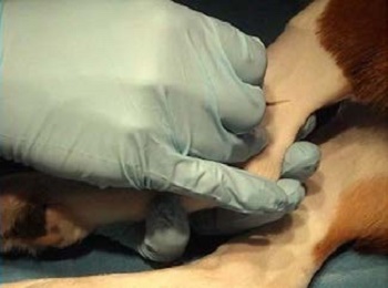
- Make a 2–3 cm incision alongside or transverse to the lateral saphenous vein.
Figure 1
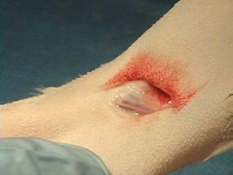
- Retract skin edges to expose the vein.
- If the vein is still distended with blood, a catheter may be inserted directly into the vein in a similar fashion to passing it through the skin.
- If the vein is collapsed or direct passage of the catheter fails, then the following technique should be used.
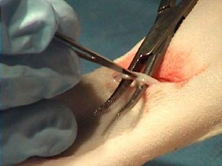
- Use curved tip mosquito forceps to bluntly strip the perivascular fascia away from the vein.
Figure 2
- With the forceps in a closed position, apply firm aggressive pressure to the fascia with the tip of the forceps.
- While maintaining pressure on the fascia, open the forceps which should strip the perivascular fascia from around the vein.
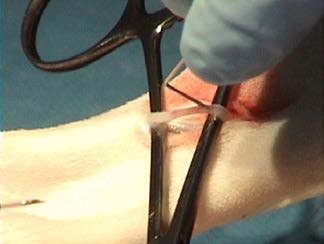
- Repeat above procedure until fascia is removed (failure to dissect sufficient perivascular tissue is a common cause of failed attempts at venous cut downs).
Figure 3
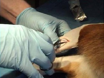
- Pass tip of forceps under the vein.
Figure 4
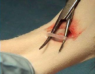
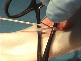
- Incise the vein with a scalpel blade—it is ok to pass a # 11 blade through both sides of the vein.
Figure 5
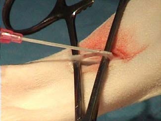
A catheter introducer can be passed into the venotomy, or a bent 22-gauge needle can be used to hold the] incision open and facilitate the passage of the catheter.
Figure 6
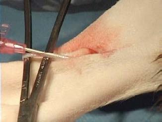
Use an over-the-needle catheter with the stylet slightly drawn back into the catheter.
Figure 7
Once the tip of the catheter is in the vein, the introducer is removed and the forceps are pulled toward the paw to straighten and stretch the vein.
Figure 8
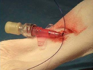
The catheter is advanced off the stylet, the stylet withdrawn, and an intravenous fluid line is connected directly to the catheter.
Figure 9
After placement, the catheter can be sutured to the vein or the wound edges can be pulled together and white tape may be used to hold the skin together and to secure the catheter in place.
Figure 10
When the animal is stable a sterile catheter should be placed in a different limb and the cut down catheter removed.
When the catheter is removed, firm pressure should be applied to the area for 2–3 minutes.
- If hemorrhage continues, the vein should be ligated.
- The skin edges can be partially closed leaving the distal portion of the incision open to drain.