The Evolution of AO/ASIF Bone Plating Equipment: Are They Better or Just Different?
Amy S. Kapatkin, DVM, MS, DACVS
Department of Surgical & Radiological Sciences, University of California, Davis
History
The date that a bone plate was first used on bone is reported to be 1565 (300 years before general anesthesia). That plate was used to repair a cleft palate and was made out of molded gold. The late 1880's brought the next major change in bone plating; surgeons began burying the bone screws below the skin. There were many designs and ideas that developed over the next 70 years. Unfortunately, malunions, nonunions and bone infections were issues due to lack of sterile techniques, and bone plates that were biomechanically unable to provide rigid fixation. Robert Danis (1880-1962) developed the ideas of compression plating and experimented with many different designs during his lifetime. Modern bone plating started in the 1950's when a group of 15 surgeons lead by Maurice Muller formed AO/ASIF (Albeitgemeinshaft fur osteosynthenfragen/ Association for the study of internal fixation) to improve the principles of bone plating. AO remains purely a medical organization to advance the study of fracture treatment while Synthes is the commercial arm of the AO.
The original plates had round holes. If compression was needed for the fracture, a separate device was needed to accomplish this. The Dynamic Compression Plate (DCP was introduced in 1969 and was the standard AO plate until a few years ago. The holes are shaped like an inclined and transverse cylinder. The screw head can slide down the incline when tightened in a vertical direction. The horizontal force of the screw head as it impacts the side of the angled hole results in movement of the bone fragment.
In an effort to balance rigid fixation and preservation of blood supply to the bone, the Limited Contact Dynamic Compression Plate (LC-DCP) was developed and released in 1990. The plate had many design features that improved the biomechanics and use of the plate such as, thinner design while maintaining equal stiffness at the screw hole s and between them, better hole design, no middle of the plate and of course the ability not to contact the periosteum in between the holes. At the same time this plate was released, surgeons were looking for methods to place plates that did not require large muscle dissection and therefore destruction of the blood supply to bone (MIPO -minimally invasive plate osteosynthesis). Systems such as the Less Invasive Stabilization System (LISS) , Point Contact Fixator (PC-Fix) and Schuhlis systems used principles of external fixation, internally and locking technology theory. What resulted in 2000 was the Locking Compression Plate (LCP) with a Combi hole so that the techniques of conventional and locked screw technology could be used in one plate.
Figure 1. The dynamic compression plate (DCP), limited contact dynamic compression plate (LC-DCP) and the locked compression plate (LCP).
The AO Mantra
The original AO principles were:
 Anatomic fracture reduction & fixation (as we know not always possible).
Rigid fracture stability (not always possible).
Preservation of blood supply through careful soft tissue approaches and fracture reduction techniques (sometimes the blood supply is damaged from the injury).
Early return to function of the plated limb (difficult in veterinary patients to control the amount of use).
With the understanding that not all fractures can be reconstructed, the "rules" have been somewhat modified to:
Long bong bones must have axial re-alignment but not necessarily anatomic perfection. Anatomic reduction is still necessary for joints.
Appropriate construct stability to ensure fracture healing via direct or indirect healing.
Atraumatic approaches and fracture reduction or minimally invasive approaches.
Early return to mobility.
Fractures can and will heal under both conditions but that is IF the appropriate condition is chosen for the appropriate fracture situation!
Conventional Bone Plating versus Locked Compression Plating
Conventional bone plates depend on direct plate to bone and screw to bone friction to maintain fracture fixation. Therefore the plates must be perfectly contoured prior to application to the bone. Fracture reduction can be lost from axial loads causing excessive shear forces on the construct that are greater than the frictional loads between the bone-plate-screw construct. The cortical screws can toggle which leads to screw loosening and loss of plate-bone fixation. Each screw works independently; the construct depends on a single screw's stiffness or pullout strength.
The biomechanical goals of the LCPs are to increase the stiffness of the construct in a biological environment. The LCP is a fixed angle construct that does not rely on screw purchase in bone. Once the screw is locked into the plate, the fixed-angle converts shear stress into compressive stress at the screw-bone interface. The load is now perpendicular to the screw axis. In order for the construct to fail under an axial load, the bone must collapse in compression. Therefore, the strength in the LCP is the sum of all the screw and plate interfaces.
Locking screws are designed with smaller threads because they are not used to generate compression between the plate and the bone. They have a larger core diameter that ensures greater bending and shear strength and dissipate the load over a larger area of bone. They have the new StarDrive head that allows 65% greater insertion torque than conventional hexagonal drivers. The StarDrive is self- retaining (stays on the screw driver without a holding device). The locked screw has a conical, double-lead thread design that facilitates alignment with the threaded plate hole.
| Figure 2. | 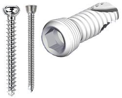
A cortical screw, a locked screw and the StarDrive head on the locked screw. |
|
| |
To date, there are no randomized clinical trials in human or animals comparing the LCP plate to conventional plates (DCP and LC-DCP) in patients with similar fractures. The plates are studied and compared in vitro (human and animal) and in case series' and are where the information on LCP principles and indications come from. The purported indications for LCPs include: patients with poor quality bone (osteoporosis, osteomyelitis), complex periarticular fracture (especially when contouring may be difficult in the metaphyseal area), inability to get minimal number of conventional screw cortices, periprosthetic fractures, when only bridge plating is possible or indicated, nonunions from failed fixations (cortex or cancellous screw stripping or screw back-out), polytrauma cases (especially when the fractures cannot be anatomically reconstructed). In vitro studies in bone models do show that locked screw constructs fail at higher loads than cortex screws and their advantage is magnified in osteoporotic bone.
Technical and biological LCP aspects that are not known when used in veterinary patients are: the ideal number of locked screws on either side of the fracture, the number of unicortical versus bicortical screws necessary for success, indications for some plate contouring (although not exact contouring), the effects of combining conventional screws and locked screws in the same construct, indications for double plating or adding additional implants (such as plate rod constructs), if there are additive biological effects on fracture healing when LCPs are placed minimally invasively. It is technically possible to place locking plates and screws minimally invasively with proper fluoroscopic equipment. In human studies there is little mechanical advantage in placing more than 2 locked screws on either side of the fracture. This may be quite different in animal patients that cannot be strictly confined or have multiple limbs fractured. Fracture fixation failures with LCPs do occur; the clinical case application will address some of the reasons for this.
References
1. Aguila AZ, Manos JM, Orlansky AS, et al: In vitro biomechanical comparison of limited contact dynamic compression plate and locking compression plate. Vet Comp Orthop Traumatol 18:220-226, 2005
2. AO Manual of Fracture Management. Internal Fixators: Concepts and Cases Using LCP and LISS. Wagner M, Frigg R (eds.), Thieme, Stuttgart, 2006
3. Egol KA, Kubiask EN, Fulkerson E, et al: Biomechanics of locked plates and screws. J Orthop Trauma 18(8): 488-493, 2004
4. Florin M, Arzdorf M, Linke B et al: Assessment of stiffness and strength of 4 different implants available for equine fracture treatment: A study on a 20° oblique long-bone fracture model using bone substitute. Vet Surg 34:231-238, 2005
5. Frigg R: Development of the locking compression plate. Injury 34:S-B6-S-B10, 2003
6. Gautier E, Sommer C: Guidelines for the clinical application of the LCP. Injury 34:S-B63-S-B76, 2003
7. Perren SM: Evolution of the internal fixation of long bone fractures. The scientific basis of biologic internal fixation: choosing a new balance between stability and biology. J Bone Jt Surg 84B:1092-1110, 2002
8. Stoffel K, Dieter U, Stachowiak G et al: Biomechanical testing of the LCP-how can stability in locked internal fixators be controlled? Injury 34: S-B-11- S-B19, 2003
9. Wagner M: General principles for the clinical use of the LCP. Injury 34: S-B31- S-B42, 2003
10. Zura RD, Browne JA: Current concepts in locked plating. J Surgical Orthop Advances 15(3): 173-176, 2006