Endemic Mycobacteriosis in Charco de Palma Pupfish, (Cyprinodon longidorsalis) Epidemiology and Effect of Antimycobacterial Therapy
Abstract
An outbreak of mycobacteriosis caused by M. marinum is described in a captive population of endangered Charco de Palma pupfish (Cyprinodon longidorsalis). This outbreak was associated with a 100% mortality over a 23-week period. Characteristic lesions of piscine mycobacteriosis were observed in all the fish examined, and mainly affected the skin (100% of the fish examined), kidney (86.7%), choroidal rete (80%), spleen (78.7%), heart (71.4%), and gills (68.2%). Despite a positive in vitro sensitivity results for the strain of Mycobacterium involved in this outbreak, a combination of rifampin and minocycline hydrochloride in the food (both at 0.3%) for 12 weeks did not decrease either the mortality rate or the severity of the lesions when compared to a untreated group. Recommendations for the control of mycobacterial infection in this species of fish include decreasing the population density, removing fish with lesions or clinical signs, separation of age classes, maintaining pupfish in fresh water instead of brackish water; and using an ultra-violet sterilization systems.
Introduction
Charco de Palma pupfish (Cyprinodon longidorsalis), a member of the Cyprinodontidae family, were once locally found in spring-fed surface waters of Northern Mexico. This species was extirpated from its natural habitat following the extensive land drainage associated with human activities. Fortunately, specimens were salvaged from these now dried up ponds, and breeding populations of this homeless fish are now maintained in several public aquaria and private collections. Charco de Palma pupfish have been successfully bred at the Toronto Zoo since 1994, but at the end of 1996, fatal cases of mycobacteriosis were diagnosed in this group. Thereafter, mycobacteriosis became endemic and highly prevalent in this population. Due to the high incidence of the disease, it was decided to interrupt the breeding program, and to evaluate the potential benefit of antimycobacterial therapy on the emergence of this infectious disease in this species of fish. This presentation has for objectives to describe the epidemiologic pattern and the significance of mycobacteriosis in captive Charco de Palma pupfish, and to evaluate the effect of an oral antibiotic treatment using a combination of minocycline hydrochloride and rifampin in the progression of this disease.
Materials & Methods
A group of 205 Charco de Palma pupfish, in which mycobacteriosis had been endemic for eight months, was used for this trial. These fish, were divided into six groups using a systematic random sampling, stratified for sex and length-classes. The three tanks of the treatment group were fed twice daily with a Toronto Zoo fish gelatin diet dosed with a combination of 0.3% of minocycline hydrochloride (Apo-minocycline, Apotex, Toronto, Canada) and 0.3% of rifampin (Rifadin, Hoechst, Montreal, Canada). The three tanks of the control groups were fed with the standard Toronto Zoo fish gelatin diet. Fish were fed twice daily with finely ground fish gelatin diet (either medicated or not) for a total of 12 weeks. Subsequently, all tanks were fed using standard, non-medicated fish flakes for the remaining 11 weeks. All tanks were checked twice daily in order to detect any dead or moribund fish. Both dead and moribund fish were immediately removed, euthanized if needed, examined for the presence of external lesions, and preserved in Bouin's (BDH Inc., Toronto, Canada). Five sagittal sections of the body, and one longitudinal section of the forehead, were embedded in paraffin wax, and 5 µm sections were stained with Ziehl-Neelsen and examined by the same pathologist (SL).
Results and Discussion
Mycobacterium marinum was isolated in pure culture from the external lesions of all four fish sampled (incubated at 18°C for two weeks on Lowenstein-Jensen-Gruft modification medium [Difco, Detroit, USA]). The isolate was sensitive in vitro to minocycline hydrochloride, and rifampin.
All fish in this trial died or were euthanized within a 23-week period. A total of 174 (90 found dead and 84 euthanatized) of the 205 initial fish were recovered, the others being most likely scavenged by tank-mates. No differences were observed in the survival curves between treated and control groups (Figure 1).
Mildly raised, white to cream, often linear, cutaneous ulcerations were observed in 68.4% of the fish. Other macroscopic findings included diffuse cutaneous edema (33.3%), exophthalmia (21.6%), and celomic distension (13.7%). The frequency and severity of these lesions did not significantly differ between treated and untreated groups.
Of the 174 fish retrieved, 17 were too autolysed to merit histologic examination. Therefore 157 fish, 76.6% of the initial fish, were examined for histology. Moderately to markedly extensive systemic lesions of mycobacteriosis were observed in 100% of the pupfish examined. Based on the severity of these lesions, we firmly believe that this infection was clinically significant in every case. Mycobacteriosis, therefore, probably accounted for most, if not all, the mortalities experienced by this population. No differences in lesion frequency (Table 1) and severity (data not presented here) were detected between the treated and untreated groups. No other significant histologic anomalies were observed in the fish examined.
Different factors might account for the very high mortality associated with this M. marinum infection. 1) Male Charco de Palma pupfish are highly energetic and territorial fish, that spend most of their time chasing each other, or displaying at females by rubbing their sides. The close and vigorous skin-to-skin contact between fish probably facilitated the spread of mycobacteria through cutaneous abrasions. These social stress might also have predisposed fish to infection by mycobacteriosis.2 2) Fish with external lesions were intentionally left in the tank until they showed systemic clinical signs. Therefore, fish with open sores were a source of infection for tank mates. 3) Contact between fish of various ages, probably increased the exposure of young naive fish to older fish with advanced stages of the disease, and might have therefore greatly increased the speed and efficacy of horizontal transmission. 4) Species of fish are not equally sensitive to mycobacteriosis,2 and some strains of Mycobacterium are more pathogenic than others.1 The Charco de Palma pupfish therefore, might be extremely sensitive to infection by M. marinum and/or were exposed to a highly virulent strain of this species.
In our study, a combination of minocycline hydrochloride (0.3%) and rifampin (0.3%) administered in the food for 12 weeks did not reduced neither the mortality rate, nor the severity of the mycobacterial lesions. The use of this therapeutic regime is therefore probably of little use for the control of infection by M. marinum in this species. Eradication of piscine mycobacteriosis in the captive population of Charco de Palma pupfish is probably unrealistic. However, different husbandry methods might help to reduce the incidence of this disease to a tolerable level. 1) Fish density should be decreased in order to minimize aggression and social stress; 2) Fish should be housed by age classes to decrease transmission from older to younger fish; 3) Contact between fry and breeding fish should also be minimized for the same reason; 4) Fish with external lesions or clinical signs should be removed to decrease the risk of transmission by predation and scavenging; 5) The use of an ultra-violet light sterilization systems may decrease the concentration of viable mycobacteria in the water, potentially decreasing the transmission rate between fish.
| Figure 1. | 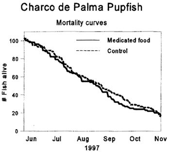
Mycobacteriosis in Charco de Palma pupfish. Comparison of mortality curves. |
|
| |
Table 1. Mycobacteriosis in Charco de Palma Pupfish.
Distribution of microscopic lesions of mycobacteriosis. Comparison between the group treated with medicated food
|
Tissues affected by microscopic lesions of mycobacteriosis |
Percentage of fish affected (%) |
[Percentage of fish affected per groupAverage for three groups (mean ± SD)] |
|
|
|
Treated group |
Control group |
|
Skin |
100 |
100.0 ± 0.0 |
100.0± 0.0 |
|
Kidney |
86.7 |
85.8 ± 10.0 |
85.6 ±5.5 |
|
Choroidal rete |
80 |
79.4 ± 10.7 |
78.7 ±6.4 |
|
Spleen |
78.6 |
85.0 ± 10.8 |
79.0 ±17.5 |
|
Gills |
68.2 |
66.1 ± 9.4 |
68.8±4.4 |
|
Celomic fat |
53.8 |
55.8 ± 12.0 |
50.7 ±7.2 |
|
Muscles and connective tissue |
52.7 |
51.2 ± 6.4 |
53.0 ± 9.2 |
|
Liver |
43.5 |
44.1 ± 14.8 |
42.7 ±10.4 |
|
Lips and oral cavity |
42.6 |
49.9 ± 14.0 |
34.7 ±3.9 |
|
Meninges |
34.9 |
31.8 ± 14.2 |
34.5 ±8.3 |
|
Ovaries |
25. |
6 38.7 ± 17.7 |
14.1 ± 6.4 |
|
Testis |
16.3 |
21.7 ± 13.4 |
8.9 ±6.8 |
|
Gastrointestinal tract |
3.8 |
6.7 ± 6.5 |
2.4 ±1.7 |
Acknowledgments
We wish to express our gratitude to the Animal Health Center keepers who participated to this project, and to the staff of the histopathology lab of the University of Guelph. Special thanks to Jeff Young and Susan Andrus for assistance and useful discussions.
References
1. Baker JA, WA Hagan. 1942. Tuberculosis of the Mexican Platyfish (Platypoecilus maculatus). Journal of Infectious diseases 70:248-252.
2. Smith SA. 1997. Mycobacterial infections in pet fish. Seminars in Avian and Exotic Pet Medicine 6:40-45.