Abstract
Rock hyrax (Procavia capensis) are unusual small mammals and appear susceptible to a range of medical problems yet are poorly represented in the veterinary literature. This paper will be based on the author’s experiences with two colonies of this species in a UK zoo over the past two decades (Table 1). Postmortem diagnoses will be described and various disease presentations discussed. In the oral presentation, sample-taking sites and a summary of “normal” hematology/biochemistry results will also be provided.
Table 1. Postmortem findings in rock hyrax at Marwell Zoo 1989–2008
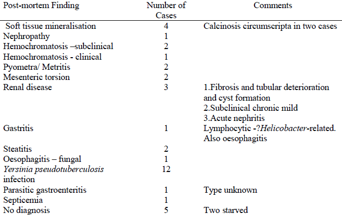
Soft Tissue Mineralization
This was a feature in three cases. In two animals, calcinosis circumscripta lesions between the toes of all feet had been investigated prior to euthanasia. A cause was not found in two cases. There was no exposure to excess Vitamin D3 either in the diet or by accidental ingestion of rodenticide. In one case there was significant renal pathology. In another there was hemochromatosis. The kidneys of this animal were too autolysed for full analysis but the nature of lesions in other organs may enable some conclusions to be drawn regarding possible renal pathology. In two cases there was evidence of renal disease, but the pathology was not felt to be severe enough to cause such lesions. Only one animal (not with calcinosis circumscripta!) showed uremia in antemortem blood sampling.
Hemochromatosis
This was found as a subclinical finding in two animals and as suspected cause of disease in one other. However, it has not been a frequent finding in these animals, and blood screening has failed to show significantly high levels of unbound iron. While testing procedures in this species are not fully evaluated, it is possible that hemochromatosis results from an individual’s faulty metabolism of iron rather than excessive dietary levels. Nonetheless further research is indicated.
Mesenteric Torsion
This was a feature in two cases of acute death. In both cases there was an underlying enteritis (presumed bacterial).
Renal Disease
In addition to the cases of soft tissue mineralization, three cases of renal disease were found. Two appeared as significant primary problems: one was believed to be subclinical.
Steatitis
Two cases were seen. In both cases vitamin E/selenium deficiencies were proposed as causes though no proof was found.
Yersiniosis
This was a major problem in the colonies over a period of 4 y. An autogenous killed vaccine was made and administered annually thereafter. Rodent control was also increased. Deaths from yersiniosis stopped at this time. Vaccination was continued for a further 6 y but has not been given for the past 6 y. No further cases have been seen even though rodents have been a problem from time to time. Blood sampling of two animals failed to find any evidence of antibody production in response to the vaccine. It is therefore not known what the decisive factor was in terms of controlling (and possibly eradicating) this infection. Lack of antibody production does not necessarily show vaccine failure and it is possible that cell-mediated immunity could be relatively long lasting. However, some animals present in the colonies now will never have been vaccinated. It is also possible that the period of rodent control and vaccination was long enough to allow the Yersinia pseudotuberculosis organism to become less prevalent in the local wild rodent population.
Parasites
It is surprising that so few internal parasites were found. Parasitic gastroenteritis was found in one animal though the parasite was not identified. An unknown lung mite was found in another. In one live animal (that presented thin) lungworm was identified on lung washes. The condition responded to anthelmintic therapy. However, lungworm has not been identified in any postmortem investigation.
Thin, Starving Animals
Two animals were identified as starved with no other findings. One was juvenile. A “wasting syndrome” is apparent in adult animals, usually in subordinate females. There are rarely any significant findings on clinical examination or on hematology/biochemistry and, apart from one case of lungworm infestation, parasitic burdens have not been found. At postmortem there are rarely significant findings other than terminal acute conditions (e.g., septicemia) or diseases that one would suspect to be related to reduced immunity (e.g., fungal oesophagitis). It is felt that the problem may be linked to group dynamics with subordinate animals being “rejected” from the colony and prevented from feeding. External wounds are rare, so it does not seem to be related to physical aggression.