Maître de Conférences, Reproduction Animale - Ecole Nationale Vétérinaire d'Alfort (Paris)
Alfort Cedex, France
It has been proven that intra-uterine artificial inseminations (AIs) in the bitch is likely to give better results, especially when using chilled or frozen semen (Linde Forsberg et al. 1995, 1999). The number of veterinarians practicing these two techniques is increasing nowadays, and we can expect that, in the coming 10 years, the exchanges of dog semen between countries will increase a lot. In some countries, however, surgical intra-uterine AIs are forbidden for ethical reasons. And when they are not, many bitch owners are reluctant to see their animal anaesthesied only to be inseminated.
To practice intra-uterine AIs, two major techniques are available.
1. Intra-Uterine Insemination Using the Scandinavian Inseminating Devices
This technique has been developed in Norway by Kjell Andersen. It is close to what is done in cattle for example. Palpating the uterine cervix through the abdominal wall, the inseminating veterinarian will try to pass a thin metallic catheter inside the opening of the cervix. This technique needs a rather long training to be confident with it, but once veterinarians are skilled in practicing it, it becomes a fast and reliable way of performing intra-uterine AIs. Only fat and giant bitches are difficult to inseminate in this way.
The inseminating devices are made of two parts: one plastic white cylindric speculum is inserted into the vagina and helps finding the cervix and guiding a metallic inseminating catheter up to the far vagina. The catheters comes in 3 different sizes: small; 20 cm. long with thinner tip for the miniature dogs, medium; 30 cm., fits most sizes, long; 40cm., for the larger dogs, (there is also an oversized which is 45 cm. long for the extreme dogs, but both the long and x-long sometimes need a stainless steel tube outside as support, otherwise they tend to bend and it is then difficult to reach the cervix for fixation).
The catheters have to be ordered in Norway (Dr Jan Fougner: fougner@norpels.no). The price for each of the three ordinary catheters is approximately in NOK (Norwegian crowns). 350. All catheters is included the plastic/nylon inserting tube. (Extra plastic speculums can only be delivered as emergency if the original is damaged).
To understand the technique, veterinarians have to remember that, in the bitch, the cervix is located at the end of a dorsal median fold, and its opening is dorsal to a big ventral dead-end (fornix) in the far vagina. Therefore, to pass the metallic catheter, the inseminating veterinarian must first push as far as possible in the vagina, and then retract it one to three centimeters behind with the right end, while the left hand grabs the cervix and turns it (pivots it) cranially so that the opening of the cervix becomes horizontal.
| Figure 1. | 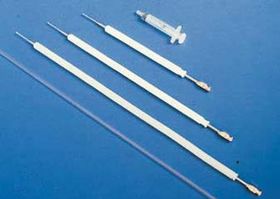
The three sizes of the Scandinavian catheters (© Linde-Forsberg) |
|
| |
| Figure 2. | 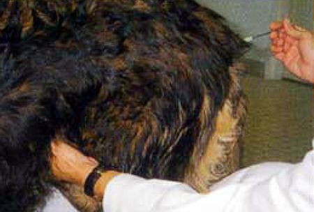
Position of the two hands of the inseminating veterinarian (© Fontbonne) |
|
| |
| Figure 3. | 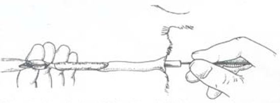
Virtual representation of the inseminating technique (Andersen 1975) |
|
| |
| Figures 4 and 5. | 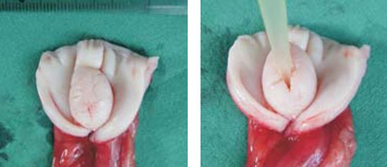
Anatomy of the far vafina, explaining the intra-uterine technique (© Fontbonne) |
|
| |
When the tip of the catheter is very close to the opening of the cervix, most of the time the operator gets a cartilaginous sensation.
Sedation is most of the time not necessary. Sometimes, when a bitch is too nervous or has a firm abdominal wall, it may be useful to sedate the animal: xylazine (Rompun®): 0.1 to 0.4 mL IV.
2. Intra-Uterine Insemination Using a Rigid Endoscope
This technique has been developed in New-Zealand by Marion Wilson. It consists of guiding an inseminating catheter (most of the time a urinary catheter--an 8 Fr is suitable but occasionally a 6 Fr may be necessary), through the cervix opening which is visualised by an endoscope often linked to a camera and to video-or computerized monitoring.
This technique needs an expensive equipment but is nearly always successful--however in some bitches it may take more than 30 minutes to manage to pass the cervix. Only miniature breeds are sometimes difficult to catheterize, as in these breeds the far vagina is often too narrow to pass the endoscope.
Endoscopic inseminations bear the great advantage of allowing the bitch owner to see what the veterinarian is doing. In this respect they may be more "commercially" valuable than the Scandinavian technique.
The requirements result from the significantly reduced space in the far vagina caused by the dorsal median fold and the particularly long vagina, which can be up to 30 cm.
Marion Wilson recommends an extended length cystourethroscope which best meets the requirements, having a 29 cm working length and 22 fr diameter. The endoscope consists of a rigid telescope with a 30 degree viewing angle together with a sheath and bridge; a cold light source and cable complete the essential equipment.
| Figure 6. | 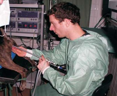
Position of the inseminating veterinarian (© Fontbonne) |
|
| |
| Figure 7. | 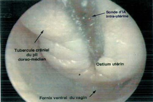
Visualization and catheterization of the cervix through the endoscope (© Fontbonne) |
|
| |
References
1. Andersen K. Insemination with frozen dog semen based on a new insemination technique. Zuchthygiene, 1975 Mar, 10(1), 1-4.
2. Linde-Forsberg C. et al. Comparison of fertility data from vaginal vs intrauterine insemination of frozen-thawed dog semen: a retrospective study. Theriogenology. 1999 Jul 1; 52(1): 11-23.
3. Linde-Forsberg C. Intra-uterine Insemination in the Dog using the Scandinavian trans-cervical catheter and a comparison with other methods. In "Recent advances in small animal reproduction" www.ivis.org, 2 Feb 2001. 6 pages.
4. Wilson M.S. Endoscopic transcervical insemination in the bitch. In "Recent advances in small animal reproduction" www.ivis.org, 12 Dec 2003. 8 pages.