There are many possible indications for extraction of teeth:
 Advanced periodontal disease
Extra (retained deciduous + supernumerary) teeth
Malformed, malpositioned + overcrowded teeth
Unerupted (impacted and embedded) teeth
Destructive (caries + resorptive) lesions
Pulp exposure, pulpitis, necrosis
Fractured teeth and jaws
Treatment failure
Other disease
In all except advanced periodontal disease the teeth are likely to remain strongly attached to the jaws making tooth removal anything but simple. Even when there has been extensive alveolar bone loss and teeth are mobile extraction can still be difficult and prone to complications such as jaw fracture.
The main problem associated with extraction is inadequate diagnosis and treatment planning so that the operator embarks on the procedure unprepared. Animals vary considerably in their anatomy, not just between species but also between breeds and even individuals of the same breed. Different teeth have different purposes and as a result different structures and they suffer from a variety of types of pathology.
The first means of simplifying extraction is to perform a thorough visual, tactile and radiographic examination so that an appropriate treatment plan can be devised to accommodate the particular anatomy of individual teeth. Once planning is complete the operating site can be prepared for the surgery by cleaning (supra and subgingival scaling, polishing and thorough oral lavage) and disinfection (application of a 0.1 to 0.2% aqueous solution of chlorhexidine) with a 3-5 minute preoperative contact time just as for any other surgery. During scaling and disinfection the appropriate sterile instrumentation can be prepared and laid out.
Once the oral cavity and surrounding skin are disinfected the animal should be positioned to allow easy access to and visualisation of the teeth requiring extraction, the surgical site then being isolated by draping.
The actual method of extraction will depend on the structure of the tooth...
Small single rooted tooth with conical root structure
Small multi-rooted tooth
Large multi-rooted tooth
Large single rooted tooth
....and its location in the oral cavity.
Most small single rooted teeth can be extracted using a "closed" extraction technique, working through the gingival sulcus without creating an access flap, although a flap often makes it easier, particularly if several adjacent teeth are to be removed. The most efficient method of separating the periodontal ligament of these small teeth is use of a thin tipped highly tapered tipped extraction instrument (a dental luxator or modern design elevator, but not a traditional chisel type dental elevator). This is used to cut the accessible portion of the periodontal ligament and stretch the alveolus to make it wider so that the tooth can be tipped within the socket (Figure 1) and the luxator inserted further.
By locking the inserted luxator against the tooth root and rotating the instrument on its long axis a large rotational leverage force (Figure 2) can be applied in a very controlled manner over a short distance. As the tooth and bone are forced apart the un-cut periodontal ligament is stretched, and if tension is held long enough, torn well beyond the tip of the instrument. By working systematically around the circumference of the root it will be loosened to the point where it can simply be lifted from its socket.
| Figure 1. Luxation technique | 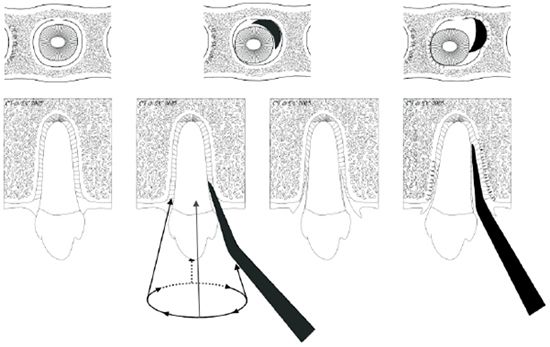
|
|
| |
If extraction forceps are used great care is required due to the long thin fragile structure of dog and cat tooth roots. This is particularly important when dealing with deciduous teeth which are proportionally longer and weaker than their permanent replacements.
| Figure 2. Rotational leverage | 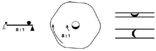
Large radius of instrument handle compared with tip width |
|
| |
By sectioning the crown using a high speed bur, after creating a shallow envelope flap, the individual roots of small multirooted teeth can be extracted in the same manner.
Large multi-rooted teeth are likely to require release of some of the bone they are attached to in order to ease extraction. These teeth should be sectioned as for smaller teeth and the access flap then enlarged to permit access for bone removal using a sterile low speed bur or bone chisels (dental elevators and luxator designs are based on bone chisels, so they are eminently suitable for cutting and removing thin and soft bone). If a bur is used a sterile isotonic irrigant (e.g., lactated ringers i/v solution) should be used to wash away bone debris and keep the bur cool. High speed dental handpieces deliver non-sterile (usually bacteria contaminated) air/water spray into the site and so are not suitable for use for cutting or removal of bone.
The buccal bone overlying the coronal half of the tooth root is released by burring around the margins or removed completely and the tooth roots loosened using luxation technique. In some cases instrument tips can also be positioned horizontally between sectioned tooth segments and rotational leverage used to tear the periodontal attachments. This method is also useful in determining whether sectioning between roots is complete; there should be some relative movement between the tooth sections as a force is applied between them.
Large single rooted teeth can be extracted similarly to individual roots of large multi-rooted teeth by gaining access, releasing the lateral alveolar wall for half of the root length and luxation of the remaining attachment. Should this not prove effective then the whole lateral wall of the alveolus can be released (Figure 3).
| Figure 3. | 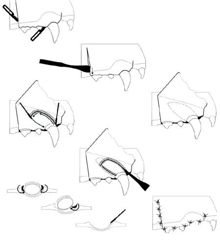
Illustration of the use of a combination of an access flap, bone release and luxation technique for extraction of the maxillary canine tooth in dogs. This method can be adapted for extraction of maxillary third incisors and mandibular canine teeth. |
|
| |
References
1. BSAVA Manual of Small Animal Dentistry (second edition). Crossley & Penman (editors). BSAVA, Cheltenham, UK. 1995.