Christian J. Wenker, DrMedVet.
Abstract
Sixteen captive plains viscachas (Lagostomus maximus), including nine animals with bilateral cataracts, were diagnosed with type II diabetes mellitus. Diagnosis was based on the presence of obesity, hyperglycemia, hypercholesteremia, elevated serum fructosamine levels, and glucosuria during a clinical examination of animals under general anesthesia. A complete diet change with marked reduction of energy was initiated, and six individuals received additional monitoring at three, six, and nine months. Mean body weight decreased by 15% over this period. Glucose and fructosamine were within reference ranges at three months, and cholesterol normalized by six months. After diet change, the captive population of plains viscachas had increased vitality and activity.
Introduction
The plains viscacha (Lagostomus maximus) is a large, colonial rodent that inhabits the pampas grasslands of Paraguay, Bolivia, and Argentina. These nocturnal herbivores live in communal burrow systems called “viscacheras” in social groups of 10–30 individuals.3 Detailed data on their current distribution are not available and they are still hunted commercially for meat and fur. As a result, this species has disappeared from most prime grasslands and agricultural areas in South America. The Zurich Zoo has bred plains viscachas regularly since 1964.8 For many years, bilateral cataract formation has been observed in neonates, which continue to worsen with age. A clinical study was initiated to ascertain whether hereditary, traumatic, biochemical, or infectious diseases were underlying factors in the development of bilateral cataracts in this species.
Methods
The plains viscachas are housed in an indoor enclosure together with a pair of burrowing owls (Speotyto cunicularia). The ground is covered with rocks, sand, and earth which provide digging activity to the animals, but additional artificial burrows of concrete tubes are also present. The diet consisted of apples, carrots, salad, bread, a commercial mineral/vitamin supplement, and rye grass hay (protein 8.9%, fat 1.7%, fiber 29.4%) with branches offered ad libitum. Sixteen animals (nine males, seven females) were anesthetized by mask induction and maintenance with isoflurane in oxygen. All animals received a complete physical and ophthalmologic examination, and the morphology of the cataract was defined in affected individuals. Blood samples were obtained from the medial branch of the saphenous vein and complete hematology counts, and serum chemistry profiles were performed. Urine was collected for urinalysis from filled urinary bladders through gentle transabdominal manual pressure or cystocentesis. Results of hematology and serum chemistry were entered into a computerized database and analyzed statistically. T-tests for independent samples were used to compare means and to evaluate significant differences between viscachas with and without cataract, and for both groups between reference values which were obtained by the author from 44 anesthetized free-ranging plains viscachas from Argentina.11
Results
All animals were obese, and nine animals showed ophthalmoscopic evidence of lens fiber and/or lens capsule degeneration, varying from focal lens opacities to severe mature cataracts. Intraocular pressure ranged from 4 to 8 mm Hg. Ocular alterations were inconclusive as to the etiology of the cataract. Glucosuria was present in four of eight animals tested. Abnormal hematologic and biochemical values (p<0.01) are listed in Table 1.
Table 1. Abnormal hematologic and biochemical values of viscachas (p<0.01)
|
Variable
|
Reference values from
free-ranging viscachas (n=44)
|
Zoo viscachas
with cataract (n=9)
|
Zoo viscachas
without cataract (n=7)
|
|
|
Mean±SDa
|
Mean±SDa
|
Mean±SDa
|
|
Glucose (mmol/L)
|
7.58±2.26
|
17.24±4.40
|
14.27±3.20
|
|
Cholesterol (mmol/L)
|
1.30±0.47
|
2.94±0.78
|
3.27±0.52
|
|
Calcium (mmol/L)
|
2.24±0.18
|
2.72±0.17
|
2.68±0.09
|
|
Fructosamine (µmol/L)
|
255.68±40.08
|
345.50±44.94
|
364.17±35.58
|
|
Lymphocytes (/µl)
|
2025±1144
|
5372±1945
|
6053±2735
|
|
Monocytes (/µl)
|
501±220
|
1232±420
|
1312±305
|
aStandard deviation.
Serum glucose values were higher in cataract-affected animals than in non-affected animals. Cholesterol, calcium, fructosamine levels, lymphocyte, and monocyte counts were lower in cataract-affected animals. However, these differences were not statistically significant between the two groups of the zoo population.
Due to significant elevations in glucose, cholesterol, and fructosamine levels with glucosuria, a presumptive clinical diagnosis of diabetes mellitus with consequent bilateral cataract formation was made. Because previous necropsies of plains viscachas with cataracts revealed immunohistochemically normal insulin production of pancreatic islet cells, the condition was classified as non-insulin dependent or type II diabetes mellitus (T2DM).
Therapy and Follow-Up
Because cataract formation was irreversible in affected animals and surgical intervention was not a feasible option, further efforts focused on the therapy of T2DM to improve the biochemical situation of the animals and prevent further cataract formation. In humans and cats with T2DM, correction of obesity by dietary measures is essential for diabetic control.2 For the captive plains viscachas, a complete diet change with marked reduction of energy and protein with an increase of fiber was instituted. Carrots, bread, apples, and salad were no longer offered. The basis of the new diet was a 1:1 rye grass hay and straw mixture which was enriched with a vitamin/mineral supplement and 15 mm herbivore pellets (Pre Alpin Lepo, Agrops GmbH, Degerndorf, Germany; protein 10%, fat 3.5%, fiber 22.7%). During the summer, fresh cut grass was offered. The new diet was well-accepted by the animals and showed a welcome behavioral enrichment in the form of greater activity.
A total of six animals, three cataract-affected and three non-affected animals, were selected for further monitoring. Anesthesia and sampling were performed as described above at three, six, and nine months after the diet change. Mean alteration of body weight was -10.7% for the zero to three-month period, -5.5% for the three to six-month period, and +1.2% for the six to nine-month period. Mean body weight decreased over the whole observation period by 15.0%. Reductions in serum glucose, fructosamine, and cholesterol is illustrated in Figures 1–3. After the diet change, glucosuria was absent. No further cataract formation occurred in previously non-affected animals and, overall, the animals showed increased vitality and activity.
Figure 1
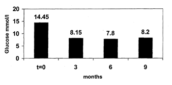
Mean long-term glucose values after change of diet in six captive plains viscachas
Figure 2
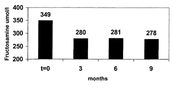
Mean long-term fructosamine values after change of diet in six captive plains viscachas
Figure 3.
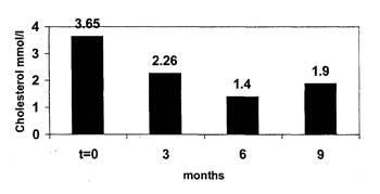
Mean long-term cholesterol values after change of diet in six captive plains viscachas
Discussion
Hyperglycemia, hypercholesteremia, and glucosuria were consistent findings for cataract-affected as well as non-affected animals, indicating a diabetic condition in the plains viscachas. However, these values can show large variability between animals, and may be influenced by food intake and stress. A glucose tolerance test after fasting was not feasible. Therefore, fructosamine levels, which have been used for diagnosis of diabetic dogs and cats and reflect glycemic control over the previous two to three weeks,7 were important to verify the diagnosis in the plains viscachas. Moderate hypercalcemia in the zoo animals may reflect the different mineral intake between the mineral-supplemented captive animals and the free-ranging reference animals. However, disturbances in calcium homeostasis are a common metabolic abnormality in diabetic humans and contribute to cataract formation.6 Lymphocytosis and monocytosis occur in chronic or acute stress situations and were related to capture and handling as well as to the diabetic condition of the animals.
Although inbreeding in the viscacha group at the zoo occurred over a long period and a genetic basis of the disease cannot be ruled out, inappropriate diet was the main factor causing T2DM and secondary cataract formation in these animals. T2DM is known to occur in several species of wild rodents in captivity if they are fed a high-energy diet.1,4,5,9,10 Most of these reported species originate in harsh, arid climates similar to the pampas. Further studies will be necessary to characterize the pathogenesis of the disease in the plains viscacha more completely, which may be a useful model for human T2DM.
Acknowledgments
This work was supported by a grant from the Committee for Zoo Animal Biology of the Zurich Zoo. I would like to thank Gabriela Hürlimann and Heinz Kohler for their indispensable help and assistance during the study.
Literature Cited
1. Barnett M, Collier GR, Zimmet P, O’Dea K. The effect of restricting energy intake on diabetes in Psammomys obesus. Int J Obesity. 1994;18:789-794.
2. Ihle SL. Nutritional therapy for diabetes mellitus. Vet Clin North Am Small Anim Pract. 1995;25:585–597.
3. Jackson JE, Branch LC, Villareal D. Lagostomus maximus. Mammalian Species. 1996;543:1–6.
4. Kalman R, Lazrovici G, Bar-on H, Ziv E. The sand rat (Psammomys obesus): morphologic, physiologic, and biochemical characteristics of a model for type-II diabetes mellitus. Contemp Topic Lab Anim Sci. 1996;35:67–70.
5. Krugner-Higby L, Shadoan M, Carlson C, et al. Type 2 diabetes mellitus, hyperlipidemia, and extremity lesions in California mice (Peromyscus californicus) fed commercial mouse diets. Comp Med Am Assoc Lab Anim Sci. 2000;50:412–418.
6. Levy J, Gavin JR, Sowers JR. DM: a disease of abnormal calcium metabolism? Am J Med. 1994;96:260–273.
7. Reusch C, Liehs MR, Hoyer M, Vochezer R. Fructosamine. A new parameter for diagnosis and metabolic control in diabetic dogs and cats. J Vet Int Med. 1993;7:177–182.
8. Rübel A, Hauser B, Ossent P. Viscachas (Lagostomus maximus), ihre Biologie, Haltung und Krankheiten im Zürcher Zoo. Verhandlungsber. Int Symp Erkr Zootiere. 1989;31:51–54.
9. Shafir E, Adler JH. Enzymatic and metabolic responses to affluent diet of two diabetes-prone species of spiny mice: Acomys cahirinus and Acomys russatus. Int J Biochem. 1983;15:1439–1446.
10. Weir BJ. The development of diabetes in the tuco-tuco (Ctenomys talarum). Proc R Soc Med. 1974;67:843–846.
11. Wenker CJ, Oppliger H, Hunziker D, Lopez J, Isenbügel E. Mission (im)possible: capture management and field research of free-ranging plains viscacha (Lagostomus maximus) in Argentina. Proc Europ Assoc Zoo Wildl Vet. 2000;3:151–154.