Mark Flückiger, PD Dr.med.vet. DECVDI
Radiographic technique
1. Minimal age for routine screening is 12 months Check specific breed-club requirements! In dogs with signs of elbow lameness radiographs should be taken at any age
2. Both elbows are radiographed
3. Rare Earth screens with a speed of 200 or less are recommended
4. No grid is used for the examination, the elbow is placed directly on the cassette
5. The beam is collimated, which improves image quality
6. The mediolateral projection is taken with the elbow in flexed position (45° opening angle) resulting in concentric superimposition of the medial and lateral humeral condyles. The MCP is best identified on a mediolateral 15° oblique view, achieved when the limb is placed in lateral position, extended and 15° supinated. Good results are achieved with a 50-60 kV-setting.
7. Additional views such as
a. mediolateral view in neutral position (approx. 110° opening angle) and
b. craniocaudal view with 15° limb pronation and 15° beam angulation in proximal direction are strongly recommended
8. Radiographs are permanently marked including the date of the examination, the identity of the dog, the identity of the owner of the dog and the clinic making the study.
Normal elbow joint, radiographs
Click on image to see a larger view
| 
mediolateral view, 45 °flexed |
|
| |
| | 
craniocaudal view, 15° pronation |
|
| |
|
Film interpretation procedure
1. Radiographs are screened for elbow disease by qualified persons. An open list of qualified persons has been filed at the FCI office by the advisory panel of the scientific committee of the FCI
2. If the elbows cannot be graded, a second examination is indicated after 3 months
3. A possibility for appeal prior to release of the results is provided
4. Results of the evaluation are open to researchers, dog owners and breeders
5. Radiographs will be archived at an appropriate location for 10 years
Film Interpretation
Radiographic findings vary depending on etiology, breed, severity, and duration of ED. The radiographic diagnosis of ED is based on presence of arthrosis and/or a primary lesion such as:
 Malformed or fragmented medial coronoid process
Ununited anconeal process
Osteochondrosis of the medial humeral condyle
Marked incongruity of the articular surface (step formation, subluxation) Further findings (of unknown etiology and relevance) may be mineralisation of periarticular tissue (flexor tendon or bursa of medial epicondyle)
DJD resulting from unknown origin
Any other abnormality noted
Radiographic findings indicative of FCP/ED
Click on image to see a larger view
Mediolateral radiograph
Increased subchondral bony density in distal part of semilunar notch, loss of trabecular pattern
Step between Radius and Ulna
Blurred cranial edge of medial coronoid process. The FCP-fragment is rarely seen!
New bone formation dorsally and laterally on the anconeal process, on the cranial border of the radius, on the medial humeral condyle, on the lateral humeral epicondyle
Uneven joint space width between humerus and radius.
Cranio-caudal radiograph
Bony irregularity and/or new bone formation on the medial border of humerus and ulna
Visualisation of bony fragments is uncommon
Step between radial and ulnar subchondral bone plate
Humeroradial joint space medial wider than lateral, particularly in BMD
Occasionally a subchondral bone defect in the medial humeral condyle with or without subchondral sclerosis (OCD or kissing lesion) is seen, but a bony flap is rare.
Beware of artifact: The sagittally running radiolucent line within the MCP usually represents the edge of the ulna but not a fissured PCM!
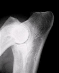
Findings with OC/OCD (Osteochondrosis, Osteochondritis dissecans)
Click on image to see a larger view
DJD similar to FCP, but usually less pronounced. Typical findings are:
Defect in articular surface of medial humeral condyle, best seen either on the craniocaudal or mediolateral extended view
A bony fragment is rarely visible
Defect may be missed when suboptimal technique is used!!
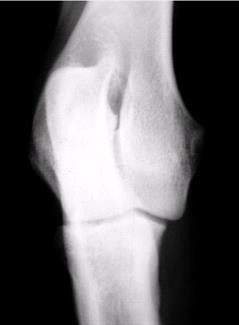
Findings with UAP (ununited anconeal process)
Irregular radiolucent vertical line between anconeal process and ulna after 18 weeks of age
Irregular subchondral sclerosis
Progressive DJD depending on duration of process
|
Click on image to see a larger view
|
Scoring
The elbow findings are scored according to severity of the arthrosis (DJD) and/or presence of a primary lesion using the IEWG (Int. Elbow Working Group) protocol
|
Elbow Dysplasia Scoring |
Radiographic Findings |
|
0 |
normal elbow joint |
normal elbow joint, no evidence of incongruency,
sclerosis or arthrosis |
|
I |
mild arthrosis |
sclerosis of ulnar trochlear notch or,
step =/> 2 mm between radius and ulna or,
osteophyte formation less than 2 mm high |
|
II |
moderate arthrosis |
osteophyte formation 2 to 5 mm high |
|
III |
severe arthrosis
or "1° ED" |
osteophyte formation more than 5 mm high or
1° ED such as UAP, FMCP, OCD |
Differential diagnoses (probably incomplete)
Common
Panosteitis (Enostosis)
Less common
Premature closure of a growth plate (usually distal ulna, traumatic in origin)
Non-traumatic short ulna syndrome or elbow malformation in chondrodysplastic dogs without elbow disease (in Basset, Corgi, and other breed)
Avulsion of flexor muscle origin at medial epicondyle
Mineralisation of flexor origins
Trauma induced elbow arthrosis
Rare
Osteomyelitis
Septic arthritis
Hypertrophic osteodystrophy
Ununited humeral condyles
Mineralisation of extensor muscle origin at lateral epicondyle
Congenital elbow luxation with lateral displacement of the radial head
Click on image to see a larger view
References
1. http://www.ncbi.nlm.nih.gov/entrez/query.fcgi (Enter: Elbow dysplasia canine)
2. Morgan JP, Wind A, Davidson A: Hereditary bone and joint diseases in the dog, schlütersche 2000.