James W. Buchanan, DVM, M Med Sci, DACVIM
The following was part of a listserve dialog on balloon valvuloplasty. It was published as a VIN page to facilitate rapid access to images.
Hello everyone,
The use of acetylcholine (ACh) to temporarily arrest the heart and facilitate balloon valvuloplasty is a reasonable approach when you are having difficulty maintaining balloon position. (A greater problem for us has been making the turn to the RV outflow tract in some small dogs.)
I have not used ACh in pulmonic stenosis cases but have had experience with it in coronary arteriography.
The attached angiograms show the effect of ACh at 0.45mg/kg with the chest open in normal experimental dogs. The 10-15 seconds arrest facilitated coronary angiography when I was developing the mitral purse string operation. Figure 1 with labels was published before medline and some of you may not have seen it. It was Figure 12 in: Buchanan JW: Selective Angiography and Angiocardiography in Dogs with Acquired Cardiovascular Disease. J. Am.Vet. Radiol. Soc. 6;5-20,1965.
Figure 1

Figure 2a shows a loose circumferential, contrast filled catheter around the mitral annulus underneath the left circumflex coronary artery.
Figure 2b shows that tightening of the catheter caused some deviation of the LCx but the artery was still patent. These images were not included in the purse string paper: Buchanan JW, Sammarco CD. Circumferential suture of the mitral annulus for correction of mitral regurgitation in dogs. Veterinary Surgery 27;182-193,1998.
Figure 2a

Cardiac arrest/aortic root injection angiogram before constricting the mitral annulus with a circumferential, contrast filled catheter.
|
Figure 2b

Cardiac arrest/aortic root injection angiogram after constricting the mitral annulus with a circumferential, contrast filled catheter.
|
Wheaton and Hamlin utilized the same ACh dose to soften the ductus and aorta during ligation of a PDA. I don't have the article handy but as I recall Dr. Wheaton mainly used it during difficult dissection and/or the few seconds it takes to tie a preplaced ligature around the PDA. I never used it for PDA ligation because I wanted to know if the ligature was tight enough to stop ductus flow at normal and increased aortic pressure which goes up when the ductus is first tied.
Regarding pulmonic stenosis (PS), I wonder if the slightly hourglass shape of an Inoue balloon or a segmental balloon for mitral stenosis would be useful in keeping the balloon in position in animals with PS. It is a bit expensive and I have not tried it.
Neumayer U, et al. Early (three-month) results of percutaneous mitral valvotomy with the Inoue balloon in 1,123 consecutive patients comparing various age groups. American Journal of Cardiology. 90(2):190-3, 2002 Jul 15.
Kasper W. et al. Percutaneous mitral balloon valvuloplasty-a comparative evaluation of two transatrial techniques. American Heart Journal. 124(6):1562-6, 1992 Dec.
The length of a balloon across the pulmonary valve is not a problem unless it is too short. Balloon size is often determined by what is available (free cast offs in the early days). The balloon used in a Samoyed with PS (Fig 3a) was sufficient to take care of any level of obstruction imaginable (Fig 3b). In my opinion, if a 4 cm won't stay in place, put in a 6 or 8 cm balloon - that will get the sucker.
Figure 3a
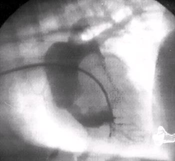
RV injection angiocardiogram demonstrating subvalvular PS, RVH and post stenotic dilation of the main pulmonary artery (MPA).
|
Figure 3b
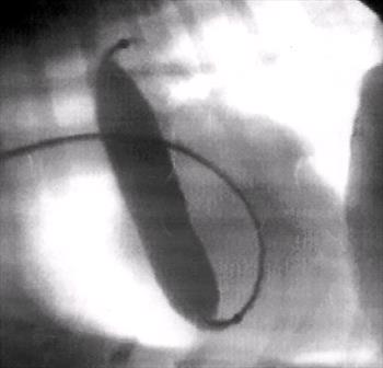
Balloon extending from the beginning of the RV outflow tract to the top of the MPA.
|
James W. Buchanan, DVM, M Med Sci
Professor Emeritus of Cardiology
Univ of Penn, Philadelphia