saddle thrombus radiograph
Arrow shows a large clot in the left atrium of a cat with a saddle thrombus. Image courtesy of Veterinary Specialists of the Valley and the North Hollywood Animal Care Center
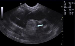
An abdominal ultrasound (previously known as a sonogram) is a diagnostic tool for looking at the organs and structures inside your pet’s belly. It can see the liver, spleen, kidneys, bladder, reproductive organs, stomach, pancreas, intestinal tract, adrenal glands, and lymph nodes. An x-ray is a still photograph, but an ultrasound is like a video.
Ultrasound uses high frequency sound waves to create live images, allowing veterinarians to get an idea of what these organs looks like and how they are functioning. The ultrasound can help discover where and why a problem is occurring so that appropriate treatment can be started. In some cases, the ultrasound findings may result in your pet needing additional testing. This does not mean that the ultrasound was unhelpful. It usually just means that the findings pointed the veterinarian in the right direction to correctly solve the pet’s problem.
Why Does my Pet Need an Ultrasound?
If your pet has been showing symptoms associated with a problem in one of those belly organs, an ultrasound may be needed. This can include diarrhea; throwing up; belly pain; abnormal urination; a mass that can be felt when pressing on the belly; or if internal injuries are suspected, such as after being hit by a car. Sometimes the veterinarian will find something specific during a pet’s physical exam, such as feeling fluid buildup in the belly, that could be seen by ultrasound. Other situations in which an ultrasound may be necessary are if lab work or x-rays show something unusual.
Abdominal ultrasounds can also be used to take samples of fluid or tissues in order to get a diagnosis of the problem. This can include guiding a needle to biopsy unusual masses or removing fluid for analysis. Veterinarians can either view these samples themselves or send them to a pathologist for testing.
As with people, ultrasounds can also be used to check for pregnancy. Pregnancy is best diagnosed after roughly 20 days from the last heat cycle. Intestinal contents and gas can occasionally cause inaccurate results. Ultrasound is not particularly useful for estimating litter or individual fetus size, so x-rays are more commonly performed. X-rays also tend to be less expensive.
The Procedure
Ultrasounds are typically done with the pet lying on a table. The veterinarian holds a transducer or probe against the belly skin. As the transducer is moved over the skin, it sends sound waves to the structures inside, which are then translated to black and white real-time images on a screen. Hair does not conduct sound waves well, so the pet’s belly is usually shaved beforehand. Alcohol or ultrasound gel may be used to provide better conduction.
Ultrasounds are usually painless and often performed in a quiet, dark room. Most pets are able to lie comfortably without stress and with minimal restraint during the brief procedure. Most pets do not need sedation. Almost all pets can safely undergo an ultrasound, regardless of medical concerns.