Environmental Aspects of Corneal Opacities in Captive South African Fur Seals (Arctocephalus pusillus pusillus)
Abstract
The associations of corneal opacities with different environmental factors were evaluated in three adult South African fur seals (Arctocephalus pusillus pusillus). The fur seals were housed in an outdoor, freshwater recirculating system using bromine as the sterilant. Between August 1995 and November 1996, the severity of the corneal opacities of both eyes was subjectively assessed daily in each animal. Potential association between the severity of these lesions and different variables were measured by Pearson’s correlation coefficients. A strong seasonal pattern was obvious for all three animals, the lesions being the most severe during the summer and the mildest during the winter. The degree of corneal opacities positively correlates with the daily maximum temperature, the global solar radiation, and the length of time with bright sunshine for all three animals. The degree of corneal opacities was poorly correlated with the length of time spent in the outdoor pool. The interpretations of these results were limited by the strong correlations between most of the variables evaluated. Even though our results did not rule out other potential etiologies, they did support the hypothesis of repetitive alterations of the corneal epithelium caused by excessive exposure to solar radiation. These alterations to the epithelial protective layer would lead to the overhydration of the corneal stroma, especially if the lesioned eye was immersed in a hypotonic fluid like fresh water. Any factors that would decrease the concentration of free-radical scavengers at the surface of the cornea, could also have a negative impact on the integrity of the corneal epithelium and could therefore enhance the formation of corneal edema.
Introduction
Corneal disease is a common health problem in captive pinnipeds.3 The exact pathogenesis of this syndrome is unclear, but circumstantial evidences suggest that different environmental factors are to blame. Proposed causal factors include osmotic stress due to low water salinity, chemical irritation caused by oxidative compounds such as chlorine and ozone, mechanical irritation associated with trauma or organic particles in suspension, nutritional deficiencies, exposure to solar radiation, and exposure to excessive light intensity.1,3,5,6
Corneal diseases of various severities have affected most of the South African fur seals (Arctocephalus pusillus pusillus) housed at the Toronto Zoo for several years. The main clinical manifestation of this syndrome is the presence of various degrees of corneal opacities. A prospective study was conducted to evaluate the temporal variations of the level of corneal opacities and to detect potential associations between the severity of these lesions and different environmental factors.
Methods
The fur seals are housed in a 25-year-old exhibit consisting of five indoor pens, an outdoor dry area, and a 50,000 gallon outdoor exhibit pool surrounded by a concrete beach with natural rocks. The pool is a freshwater recirculating system equipped with a vacuum diatomaceous earth filter with a turnover rate of approximately 2 hours. Bromine is continuously added to the system as the sterilant and the pH has been maintained around 7.6 during the time of the study. The temperature of the water freely fluctuates throughout the year from 1–25°C. The diet of these fur seals is primarily composed of Pacific herring (Clupea harangus), capelin (Mallotus villosus) and Californian squid (Illex spp.). Daily supplementation includes 6 g of NaCl, 50 mg of thiamin, 200 IU of vitamin E, and 1000 IU of vitamin C per animal.
This study was conducted between August 1995 and November 1996 on the three youngest adult fur seals of the herd, a 7-year-old male, a 7-year-old female, and a 9-year-old female. The severity of the corneal opacities of both eyes was subjectively assessed daily in each animal using the following grades: 0=No visible opacity; 1=Slight cloudiness of the cornea; 2=Moderate cloudiness of the cornea; 3=Severe cloudiness of the cornea. This evaluation was conducted by a limited number of observers and is believed to be a reasonable estimate of the severity of the corneal opacities. The length of time spent by each animal in the outdoor exhibit pool was recorded for each day. Environment Canada provided the daily maximum temperature, the daily global solar radiation, and the daily length of time with bright sunshine for the region of Toronto. The global solar radiation is defined as the total incoming direct and diffuse short wave solar radiation on a horizontal surface (MJ/m2). The length of time with bright sunshine is a measure of light intensity and is defined as the period of time with sunshine of sufficient intensity to scorch or burn a standard sunshine card installed in a Campbell-Stokes Sunshine Recorder. Weekly averages were calculated for each of the variables and were used for all graphic representations and statistical analysis. The strengths of association between the severity of the corneal opacities and the different variables were measured by Pearson’s correlation coefficients. A significance level of p=0.05 was used in all statistical tests. Statistical analyses were performed using Statview software (Abacus Concepts, Inc, Berkley, CA, 1996).
Results
The male fur seal was more severely affected than the females, but the degree of severity was significantly correlated between animals (from 0.62 to 0.75). A strong seasonal pattern was obvious for all three animals, the lesions being the most severe during the summer and the mildest during the winter (Figs. 1, 2). The degree of corneal opacities positively correlates with the daily maximum temperature, the global solar radiation, and the length of time with bright sunshine for all three animals (Table 1). These correlation coefficients did not significantly differ from each other (data not presented). A weak, but statistically significant, correlation was present between the degree of corneal opacities and the time spent in the outdoor pool exhibit for only one of the three fur seals.
Correlations were also detected between the daily maximum temperature and the global solar radiation (r=0.71; p<0.0001), the daily maximum temperature and the length of time with bright sunshine (r=0.58; p<0.0001), and the global solar radiation and the length of time with bright sunshine (r=0.88; p<0.0001).
Table 1. Correlation coefficients (r) between the severity of the corneal opacities and different variables in three South African fur seals
|
|
Male
|
Female
|
Female
|
|
r
|
p-values
|
r
|
p-values
|
r
|
p-values
|
|
Time spent in pool exhibit
|
0.17
|
0.23
|
0.36
|
0.007a
|
0.04
|
0.78
|
|
Daily maximum temperature
|
0.59
|
<0.0001a
|
0.71
|
< 0.0001a
|
0.55
|
< 0.0001a
|
|
Daily global solar radiation
|
0.6
|
< 0.0001a
|
0.69
|
< 0.0001a
|
0.68
|
< 0.0001a
|
|
Daily bright sunshine
|
0.45
|
0.0009
|
0.56
|
< 0.0001a
|
0.51
|
< 0.0001a
|
aStatistically significant.
Figure 1
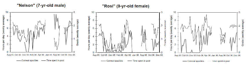
Temporal variations in the degree of corneal opacities and the length of time spent in the outdoor pool exhibit for each South African fur seal.
Figure 2
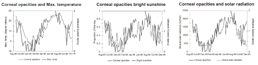
Temporal variation in the degree of corneal opacities among three South African fur seals, and the daily maximum temperature, the length of time with bright sunshine, and the global solar radiation for Toronto, Canada.
Discussion
The strong seasonal pattern observed in this study for corneal opacities suggest that the exposure to the causal factor(s) vary throughout the year. Even though the severity of the corneal opacities was different between animals, the temporal variation in the degree of severity was similar. As a result the lesions present in the three animals are probably caused by the same exposure factor(s) but with individual variation of susceptibility.
In terrestrial animals, corneal opacities are mainly associated with corneal edema, which commonly result from a disruption in the corneal endothelial pump function.2 A similar pathogenesis was proposed to explain the presence of corneal edema in pinnipeds by Bellhorn1 who speculated that the extreme myosis consequent of exposure to high light intensity was associated with repetitive damages to the corneal endothelium following the contact between the constricted iris and the endothelial surface.1 As a result of this damage, the disruption of the osmotic pump would lead to overhydration of the corneal stroma.1 However, this hypothesis was not supported by subsequent detailed ophthalmologic examinations of affected and non-affected pinnipeds.6 In our study, a positive correlation was seen between the severity of corneal opacities and the length of time with bright sunshine (a measure of light intensity). Even though this finding would support Bellhorn’s hypothesis, the apparent correlation could also be due to other confounding factors, such as global solar radiation, and ambient temperature that were also highly correlated to this variable. Subsequently, if the exposure to bright sunshine was the main factor in the development of these corneal opacities, higher degrees of corneal opacities would have been expected to occur during some of the sunny periods of winter, however this was not the case.
Epidemiologic evidence in California sea lions (Zalophus californianus),3 and several anecdotal reports suggests that the exposure to fresh water is an important factor that contributes to the development of corneal edema in pinnipeds. However, due to the strong resistance of the corneal epithelium of pinnipeds to osmotic pressure, and to the fact that pinnipeds kept in fresh water are frequently free of corneal lesion, low salinity is not believed to be a primary causal factor.6 Our findings are in agreement with this statement (Table 1). However, the absence of strong correlation between the time spent in the outdoor exhibit pool and the severity of the lesions should be interpreted with caution due to possible bias associated with the management of corneal lesions in these animals. Fur seals with severe opacities were frequently kept either indoor or in the dry area until improvement of the ocular condition. This management practice might have hidden a potential association between this syndrome and the exposure to fresh water. Nevertheless, we proposed that the exposure to fresh water is probably not a primary cause, but instead an aggravating factor that needs to be preceded by alteration to the corneal epithelium. Overhydration of the corneal tissue (edema) would occur following a dysfunction of the corneal epithelium and would be aggravated by the contact with the hypotonic external milieu that constitutes fresh water. Accordingly, the hypertonicity of the salt water would offer some protection.
The integrity of the corneal epithelium can be altered by different environmental factors. Irritations caused by particles in suspension, trauma, or chemicals used for disinfection can potentially induce inflammatory or mechanical changes that could affect the integrity of the corneal epithelium.5 The results of our study do not support the role of a water-borne irritant as the primary cause of the lesions observed in our group of fur seals (Table 1). However, as for the exposure to fresh water, a possible bias associated with the management of this syndrome exists and limits the interpretation of this finding. Exposure to solar radiation has been proposed as a factor that can affect the integrity of the corneal epithelium.6 Through different mechanisms, such as the formation of free radicals, exposure to ultraviolet light is known to alter the cellular permeability of epithelial cells. The strong seasonal pattern observed in our study and the positive correlation between the severity of the lesions and the level of global solar radiation for all three animals in our study support this hypothesis. However, as for the exposure to bright sunshine, the possibility of confounding factors cannot be ruled out with our data. Interestingly, syndromes causing corneal opacities have been linked to chronic exposure to solar radiation in humans.4 In these syndromes (climatic droplet keratopathy, and pterygium), the corneal opacities are actually not consequent to edema, but to the accumulation of precipitated proteins or to an increase in fibroblasts and hyalinization of connective tissue.4 These conditions might share some similarities with the chronic corneal opacities encountered in pinnipeds, and deserve further investigations.
The main limitation of this prospective study is the strong correlations between most of the variables evaluated. Because of this, the effects of confounding variables cannot be controlled for. It is also not possible to rule out other factors that were not evaluated in this study, like variation in water chemistry, and the effects of exhibit design. Nevertheless, we believe that without ruling out other potential etiologies, our study supports the hypothesis of repetitive alterations of the corneal epithelium caused by excessive exposure to solar radiation. Other irritants, such as particles in suspension and bacterial aggressions might also, by inducing inflammatory or mechanical changes, contribute to alter the composition of the corneal epithelium. These alterations to the epithelial protective layer will lead to the overhydration of the corneal stroma, especially if the eye lesion is immersed in a hypotonic fluid like fresh water. Any factors that can decrease the concentration of free-radical scavengers at the surface of the cornea, including nutritional deficiencies and aging, could also have a negative impact on the integrity of the corneal epithelium and could therefore enhance the formation of corneal edema.
Acknowledgments
We are grateful to the keepers of the South African Fur seals of the Toronto Zoo and to Margaret Reed for her help with data entry.
Literature Cited
1. Bellhorn, R. W. 1977. Corneal opacities in marine mammals: A light induced phenomenon. Eighth Annual Conference and Workshop of the International Association for Aquatic Animal Medicine. Boston, Massachusetts.
2. Dohlan, C. H. 1994. Corneal Edema. In: Alber, D. M. and F. A. Jakobiec (eds.). Principles and Practice of Ophathalmology. W. B. Saunders Company, Philadelphia, Pennsylvania. Pp. 245–256.
3. Dunn, J. L., N. E. Overstrom, and D. J. St.Aubin. 1996. An epidemiologic survey to determine factors associated with corneal and lenticular lesions in captive harbor seals and California sea lions. Proceeding of the International Association for Aquatic Animal Medicine. Chattanooga, Tennessee. p. 100.
4. Klauss, V. K. and E. C. Schwartz. 1998. Other conditions of the outer eye. In: Johnson, G. J., D. C. Minassian, and R. Weale (eds.). The Epidemiology of Eye Disease. Lippincott-Raven Publishers, Philadelphia, Pennsylvania. Pp. 137–158.
5. Rigdeway, S. H., J. R. Geraci, and W. Medway. 1975. Diseases of pinnipeds. Rapp. P. V. Reun. Cons. Int. Explor. Mer 169:327–337.
6. Stoskopf, M. K., L. W. Hirst, and D. Graham. 1983. Ocular anterior segment disease in captive pinnipeds. Aquatic Mammals 10:34–44.