Scott P. Terrell1,2, DVM, DACVP
Abstract
In December 2001 and January 2002, more than 3000 turtles entered the United States as part of the efforts of the Turtle Survival Alliance (TSA) and Kadoorie Farm and Botanical Garden (KFBG) to save illegally harvested wild Asian turtles. The turtles were part of a large group of approximately 7500 turtles that had been confiscated by Hong Kong officials in December 2001. Veterinarians, biologists, conservationists, turtle enthusiasts, and other volunteers assisted in the triage, treatment, and transport of these turtles as they arrived at a private facility in central Florida. Many of the turtles arrived severely debilitated or dead. Necropsies were performed either on-site, during triage efforts, or at a separate facility. Preliminary results of gross pathology and bacterial culture (Table 1) for 65 turtles are presented. Antimicrobial sensitivity results are summarized in Table 2.
Table 1. Results of bacterial cultures from four species of Asian turtles
|
Turtle species
|
Liver primary organism
|
Liver secondary organism
|
Lung primary organism
|
Lung secondary organism
|
|
S. crassicollis
|
A. hydrophila
|
Gram negativea
|
Gram negativea
|
Proteus sp.
|
|
H. grandis
|
A hydrophila
|
Acinetobacter sp.
|
A. hydrophilab
|
|
|
“
|
A. hydrophilab
|
|
A. hydrophilab
|
|
|
“
|
A. hydrophilab
|
|
A. hydrophilab
|
|
|
“
|
A. hydrophilab
|
|
A. hydrophila
|
Alpha Strepc
|
|
“
|
A. hydrophilab
|
|
A. hydrophila
|
E. coli
|
|
“
|
A. hydrophilab
|
|
A. hydrophilab
|
|
|
“
|
A. hydrophilab
|
|
Providencia sp.
|
A. hydrophila
|
|
O. borneensis
|
A. hydrophila
|
Proteus sp.
|
Gram negativea
|
A. hydrophilab
|
|
“
|
A. hydrophila
|
Proteus sp.
|
Not cultured
|
|
|
“
|
A. hydrophilab
|
|
A. hydrophila
|
Proteus sp.
|
|
“
|
A. hydrophilab
|
|
A. hydrophila
|
Proteus sp.
|
|
“
|
Gram negativea,b
|
|
A. hydrophila
|
Proteus sp.
|
|
“
|
Gram negativea
|
E. coli
|
A. hydrophila
|
Proteus sp.
|
|
“
|
A. hydrophilab
|
|
Serratia sp.
|
A. hydrophila
|
|
“
|
A. hydrophila
|
Proteus sp.
|
A. hydrophila
|
Gram negativea
|
|
“
|
A. hydrophila
|
Proteus sp.
|
A. hydrophila
|
Gram negativea
|
|
“
|
A. hydrophila
|
Gram negativea,b
|
Not cultured
|
|
|
“
|
Gram negativea,b
|
|
Gram negativea
|
Klebsiella pneumoniae
|
|
M. emys
|
A. hydrophila
|
Gamma Strepd
|
A. hydrophila
|
Gamma Strepd
|
|
“
|
Citrobacter freundi
|
Gamma Strepd
|
A. hydrophila
|
Citrobacter freundi
|
|
“
|
A. hydrophila
|
Pseudomonas aeruginosa
|
A. hydrophila
|
Flaviomonas sp.
|
aPure culture.
bUnidentified gram-negative bacillus.
cAlpha hemolytic Streptococcus sp.
dGamma hemolytic Streptococcus sp.
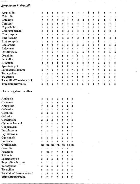
Table 2. Antibiotic sensitivity results for bacterial isolates from affected turtles. Each column represents a separate bacterial isolate.
Sixty-five turtles and tortoises representing seven different species were necropsied. The species and number examined included 28 Bornean black leaf turtles (Siebenrockiella crassicollis), 15 Malaysian giant turtles (Orlitia borneensis), 10 giant Asian pond turtles (Heosemys grandis), 6 Malayan box turtles (Cuora amboinensis), 3 Asian brown tortoises (Manouria emys), 2 leaf turtles (Cyclemys sp.), and 2 spiny turtles (Heosemys spinosa).
S. crassicollis
Twenty-eight turtles (nine males, nineteen females) were examined. One female was gravid and contained two eggs. Most (21/28) were in poor body condition based on the absence of, or severe serous atrophy of, intracoelomic fat. The coelomic cavities of 17 turtles contained fluid interpreted as ascites, possibly associated with hypoproteinemia. The stomachs of all 28 were empty and the intestinal tract contained scant dry feces. Small nematodes were present in the distal intestinal tract of two turtles. Pneumonia was evident grossly in 10/29. A fishhook was found protruding from the anus of one turtle but was not associated with any gross intestinal pathology. Aerobic culture of the liver and lung was performed on one animal (Table 1). The preliminary cause of death in the majority of this species was severe emaciation and bacterial pneumonia.
O. borneensis
Fifteen turtles (seven males, eight females) were examined. All 15 turtles were in poor body condition. The coelomic cavities of nine contained ascitic fluid. The stomachs of all turtles were empty. There were numerous large seeds or tubers present within the distal intestine that distended and often impacted the tract. The seeds were submitted to a botanist for identification (pending). The most striking finding in this species was the presence of fishhooks in the esophagus and periesophageal connective tissue. In five turtles, fishhooks were associated with esophageal perforation and a fibrinocaseous exudate. Pneumonia was present in 9/15. Other gross findings included myocarditis, splenitis, hepatitis, and coelomitis. Aerobic culture of the lung and liver was performed on 11 turtles. The preliminary cause of death in the majority of this species was determined to be bacterial septicemia and pneumonia with numerous fishhook injuries.
H. grandis
Ten turtles (five males, five females) were examined. Two females were gravid containing five and six eggs, respectively. Three turtles were severely autolyzed. Nine were in poor body condition. The stomach and intestine of all 10 turtles were filled with fluid. One turtle had massive numbers of nematodes in the distal intestinal tract. Severe hepatic lipidosis was present in four of ten; the remaining six turtles had varying degrees of lipid accumulation in the liver. There was no gross evidence of pneumonia or other inflammatory lesions in any of the turtles examined. Aerobic culture of the lung and liver was performed on seven turtles. The preliminary cause of death of the majority of this species was determined to be severe emaciation.
C. amboinensis
Six turtles (four males, two females) were examined. One female was gravid containing six eggs. All six turtles were in poor body condition. The stomach and intestine of all six were empty. Bacterial cultures were not performed on this species. The preliminary cause of death of these turtles was determined to be emaciation.
M. emys
Three female turtles were examined. None of these turtles was gravid. Two were considered to be in poor body condition. The third turtle was in moderate body condition having been fed via a percutaneous esophageal feeding tube. All three animals had been tube fed prior death and the stomachs contained abundant moist ingesta. Massive numbers of nematodes were present within the intestine of two animals. In these turtles, parasites were so numerous that they distended and impacted the mid-intestinal tract. In one turtle numerous cross-sections of parasites could be seen within the submucosa of the intestine on cut section. The embedded parasites were often associated with a caseous exudate. A diffuse severe fibrinonecrotic enteritis was evident upon removal of the parasites. Aerobic culture of the lung and liver was performed on all three turtles. Enteric pathogen culture was performed on the intestinal contents of one turtle. The preliminary cause of death of these two turtles was severe endoparasitism resulting in emaciation, enteritis, and probable bacterial septicemia.
Cyclemys sp.
Two turtles (one male, one female) were examined. The female was not gravid. Both animals were in poor body condition, and had shell defects at the bridge between the plastron and carapace, consistent with crushing. Both turtles had severe localized coelomitis associated with the bridge lesions. One turtle had grossly evident pneumonia. Bacterial cultures were not performed. The preliminary cause of death of these two turtles was traumatic shell damage resulting in coelomitis and presumptive bacterial septicemia.
H. spinosa
Two female turtles were examined. One was in poor body condition. The other was in excellent body condition with abundant fat stores. The stomach and intestine of these turtles was empty. Abundant ascitic fluid was present in the coelom. No other gross lesions were noted. Bacterial cultures were not performed. No preliminary cause of death was determined for these animals.
Postmortem examination of these turtles has provided a wealth of information about the causes of morbidity and mortality in these animals. Gross necropsy findings were relayed to clinical veterinarians in a timely fashion during triage and treatment efforts to guide therapy. Most of the turtles succumbed to emaciation and bacterial pneumonia/septicemia. Other conditions identified included fishhook injuries, traumatic injuries, endoparasitism, and enteritis. Bacterial culture and antimicrobial sensitivity results were received and relayed to clinical veterinarians approximately 1 wk after completion of initial triage and treatment and were valuable in guiding treatment for those turtles that remained in captivity. Antimicrobial sensitivity data will also be valuable in the event of future confiscations.
Further evaluation of tissues and samples collected from these turtles are on-going and include histopathology, parasite identification, examination of intestinal contents by botanists (specifically the seeds found in the intestinal tract of O. borneensis), as well as other ancillary diagnostic testing.