Abstract
Weedy sea dragons (Phyllopteryx taeniolatus) are related to seahorses and pipefish, and are found in reefs and sandy underwater regions of Western Australia, South Australia and further east along the coastline of Victoria, Australia. These fish are protected under Australian law and have become popular recently for aquarium exhibits in the United States. With the exception of a few parasites,1-3 little is known of the pathogens of sea dragons. Myxozoanosis has not previously been reported in sea dragons. This report describes the occurrence of renal myxozoanosis in 11 weedy sea dragons.
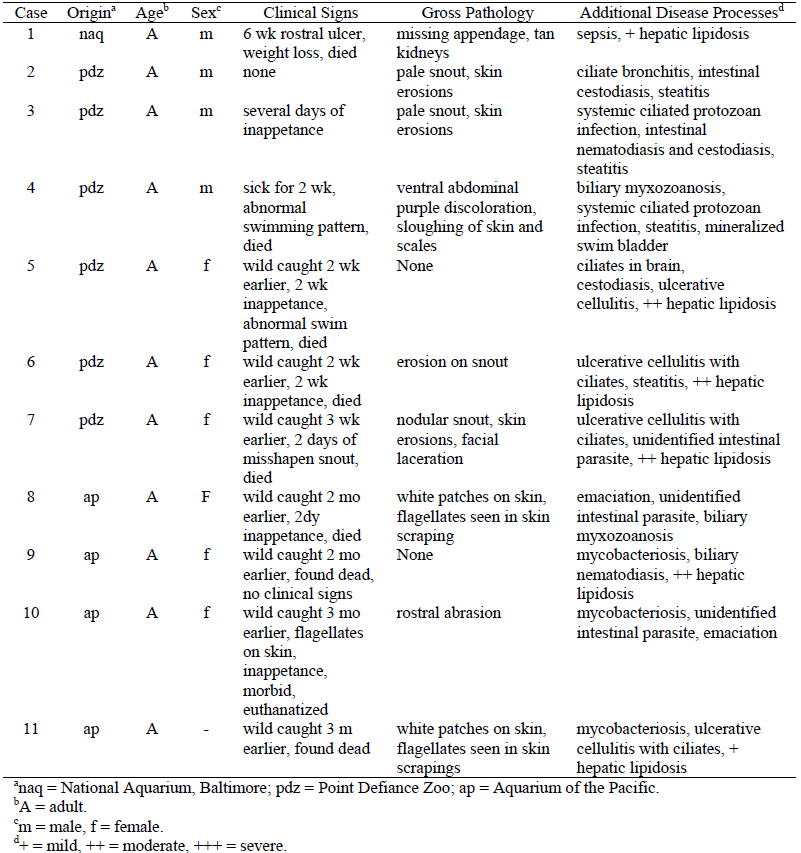
Results of this study are summarized in Table 1. The sea dragons with renal myxozoanosis were from three separate aquaria in the United States, and all cases were diagnosed during May–June of 2003, except one case that was diagnosed in March 2002. All were wild caught, and had been in captivity at the National Aquarium, Baltimore, MD (case 1), Point Defiance Zoo, WA (cases 2–7), and Aquarium of the Pacific, CA (cases 8–11). All were adults of unknown age. Six were female, four were male, and one was undetermined. Inappetence was the most common clinical sign. Skin erosions and snout abrasions or deformities were the most common gross lesions. Important concurrent disease processes included cutaneous and/or systemic ciliated protozoan infection (seven cases), hepatic lipidosis (six cases), steatitis (four cases), mycobacteriosis (three cases), and biliary myxozoanosis (two cases).
Table 1. Signalment, history and clinical signs, gross and microscopic pathology of weedy sea dragons (Phyllopteryx taeniolatus) with renal myxozoanosis

Histologically, all animals had large numbers of developing myxosporeans in the lumina of renal tubules and collecting ducts. In all cases, this finding was associated with a mild degree of renal tubular dilation, hypertrophy of tubular epithelial cells, and accumulations of cellular debris and some proteinaceous fluid in the tubules.
Myxozoan spores in wet mounts and histologic sections had morphology consistent with that of the genus Sinuolinea.4 Members of this genus have been described from the urinary bladder of a variety of marine fishes. Spores were characterized from one specimen as spherical or sub spherical (16.7 µm length x 16.5 µm width) with two spore valves joined by a sutural line that curved around the spore, forming a well-marked sutural ridge. The two polar capsules (5.4 x 5.2 µm) located anteriorly were set widely apart and contained polar filaments with approximately six turns. Analysis of the 18S rDNA gene will help clarify the taxonomic position of this isolate.
The significance of renal myxozoanosis to the health status of captive weedy sea dragons is not known. All fish had one or more concurrent disease processes that could account for morbidity and/or mortality. Morphologic alterations associated with the presence of these parasites in the renal tubules were generally mild, although the impact of this infection on renal function was not determined. Antemortem detection of this parasite may be possible, as spores are shed into the urine. Because all of the fish were recent captives, it is possible that this infection occurs naturally in wild weedy sea dragons. Further characterization of the parasite may provide additional information regarding its life cycle, and the potential for spread to other fish on exhibit.
Literature Cited
1. Umehara A, Y Kosuga, H Hirose. 2003. Scuticociliata infection in the weedy sea dragon Phyllopteryx taeniolatus. Parasitol. Int. 52: 165–8.
2. Upton SJ, MA Stamper, AL Osborn, SL Mumford, L Zwick, MJ Kinsel, RM Overstreet. 2000. A new species of Eimeria (Apicomplexa, Eimeriidae) from the weedy sea dragon Phyllopteryx taeniolatus (Osteichthyes: Syngnathidae). Dis. Aquat. Org. 43: 55–9.
3. Langdon JS, K Elliott, B MacKay. 1991. Epitheliocystis in the leafy sea-dragon. Aust. Vet. J. 68:244.
4. Lom J, I Dyková. 1992. Protozoan Parasites of Fishes. Elsevier, New York, Pp. 159–235.